
|
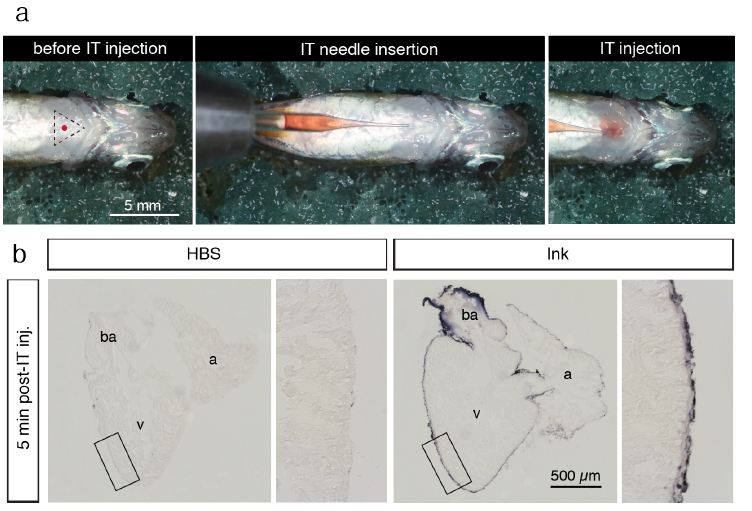
Fig. S6
Intrathoracic microinjection procedure. (a) Photographs of the microinjection procedure under the stereomicroscope. Before intrathoracic (IT) injection, an anesthetized fish was positioned with the ventral side up on a moist sponge. The tip of the pulled glass needle with a 2.5 μl solution was inserted under the skin of the thorax. The solution was slowly injected into the pericardial activity with a caution not to puncture the heart. (b) Histological sections of the zebrafish heart for a validation of accuracy of the microinjection procedure. The fish were euthanized and IT-injection was performed using 2.5 μl Hank’s buffer (HBS) or purple ink. At 5 min post injection, the hearts were collected, fixed and sectioned. Injected ink labeled the surface of the ventricle (v), atrium (a) and bulbus arteriosus (ba), suggesting efficient spreading of the injected solution throughout the pericardial cavity. No dye was observed inside the organ, demonstrating that the needle did not puncture the heart during injection.