Fig. 1
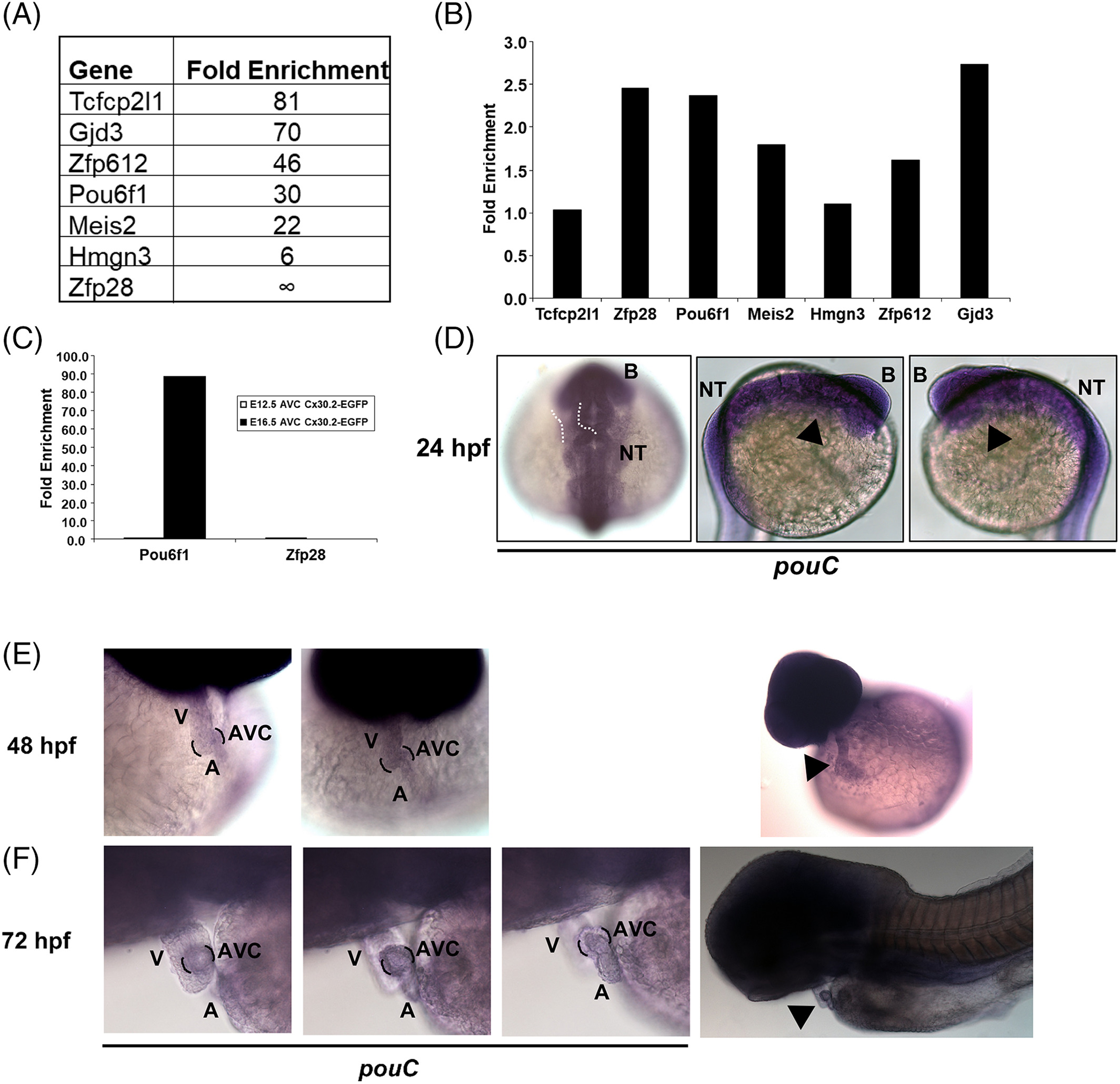
Mouse Pou61 and zebrafish pouC are expressed in the AVC. A: Cx30.2?lacZ+ mouse AVC cells were isolated at E12.5 by flow cytometry and subsequently profiled by microarray analysis (Harris et al., 2014). The top six transcription factors based on fold enrichment in Cx30.2?lacZ+vs. Cx30.2?lacZ? AVC cells are shown along with Gjd3/Cx30.2 for comparison. B: The AVC of E10.5 hearts were micro?dissected and used to generate cDNA for qRT?PCR analysis with the indicated primers. Fold enrichment is shown relative to E10.5 whole heart and normalized to Gapdh. C: E12.5 and E16.5 AVC tissue was micro?dissected from Cx30.2?EGFP transgenic mice, and GFP+ and GFP? cells were isolated by flow cytometry. RNA from each cell population was used to created cDNA for qRT?PCR analysis with the indicated primers. Fold enrichment refers to GFP+ vs. GFP? populations and normalized to Gapdh. D: In situ hybridization of a 24 hpf embryo using a pouC riboprobe. Dashed white outline demonstrates expected location of the heart at this developmental stage. Black arrowhead indicates location of the heart. E,F: In situ hybridization with a pouC probe was performed in zebrafish at 48 (E) and 72 (F) hpf. Wide angle images are shown at right for perspective. Black brackets indicated AVC region, while black arrowhead highlights pouC expression in the heart. B, brain; NT, neural tube; V, ventricle; A, atrium.

