Image
Figure Caption
Fig. S5
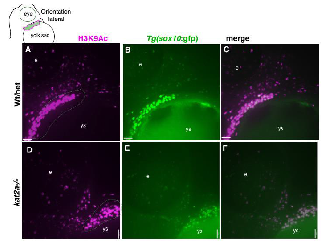
H3K9Ac is reduced in kat2a-/- mutants. Confocal micrograph for antibody staining of H3K9Ac (A, D), sox10:GFP (B, E) in wt (A?C) and kat2a-/- (D?F) whole-mount zebrafish embryos at 48 hpf, in lateral views oriented as indicated in schematic. e, eye, ys, yolk sac. Scale bars represent 100 ?m.
Acknowledgments
This image is the copyrighted work of the attributed author or publisher, and
ZFIN has permission only to display this image to its users.
Additional permissions should be obtained from the applicable author or publisher of the image.
Full text @ J Dev Biol

