Fig. 1
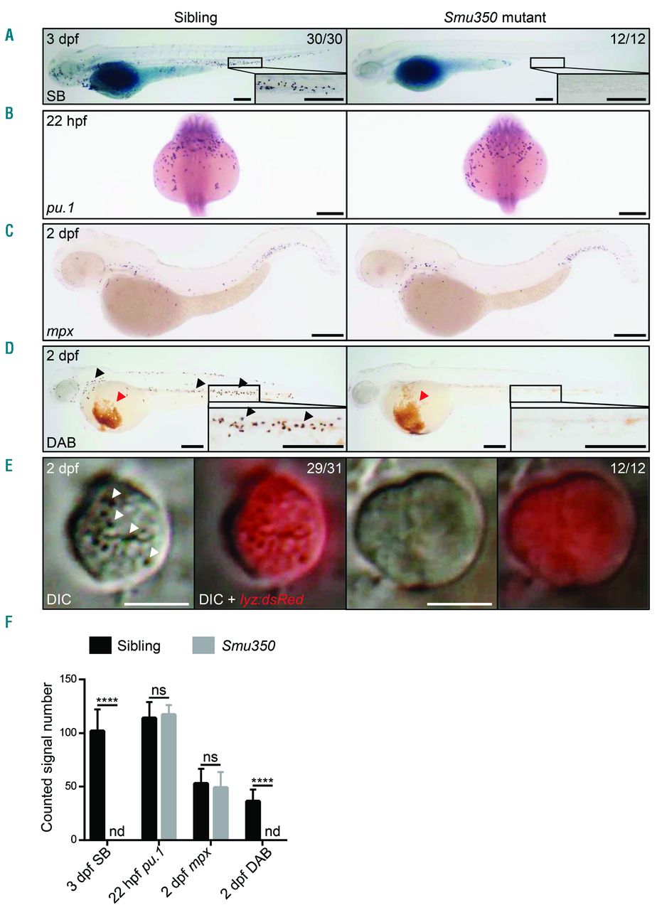
Neutrophil deficiency in smu350 mutants. (A) The Sudan black B (SB) signal was totally absent in smu350 mutants. SB staining in siblings (left) and smu350 mutants (right) at 3 days post fertilization (dpf). The boxed regions are magnified in the lower right-hand corner. (B) Whole-mount in situ hybridization (WISH) of pu.1 expression in siblings (left) and smu350 mutants (right) at 22 hours post fertilization (hpf). (C) WISH of mpx expression in siblings (left) and smu350 mutants (right) at 2 dpf. (D) DAB staining in siblings (left) and smu350 mutants (right) at 2 dpf. Signal points representing neutrophil peroxidase activity (black arrowheads) were absent in smu350 mutants. Red arrowheads indicate the hemoglobin signal. The boxed regions are magnified in the lower right-hand corner. (E) Neutrophil granules were absent in smu350 mutants. In vivo imaging of neutrophils in 2-dpf Tg(lyz:DsRed);smu350 embryos by VE-DIC microscopy. Neutrophils of siblings had abundant visible and highly mobilized granules (29 of 31 embryos), while neutrophils of smu350 mutants lacked granules (12 of 12 embryos). White arrowheads indicate neutrophil granules. (Left) Bright-field DIC image; (right) an overlay of bright-field DIC and fluorescent images. See also Online Supplementary Appendix, Movies 1 and 2. (F) Quantifications of SB+ cells in the posterior blood island (PBI), pu.1+ cells (B), mpx+ cells in the PBI (C), and DAB+ cells in the PBI (D). MeanąStandard Deviation (SD), n>15; Student t-test: ****P<0.0001. ns: not significant; nd: not detectable. Scale bars: 200 ?m (A-D) and 5 ?m (E).

