
|
Fig. 2
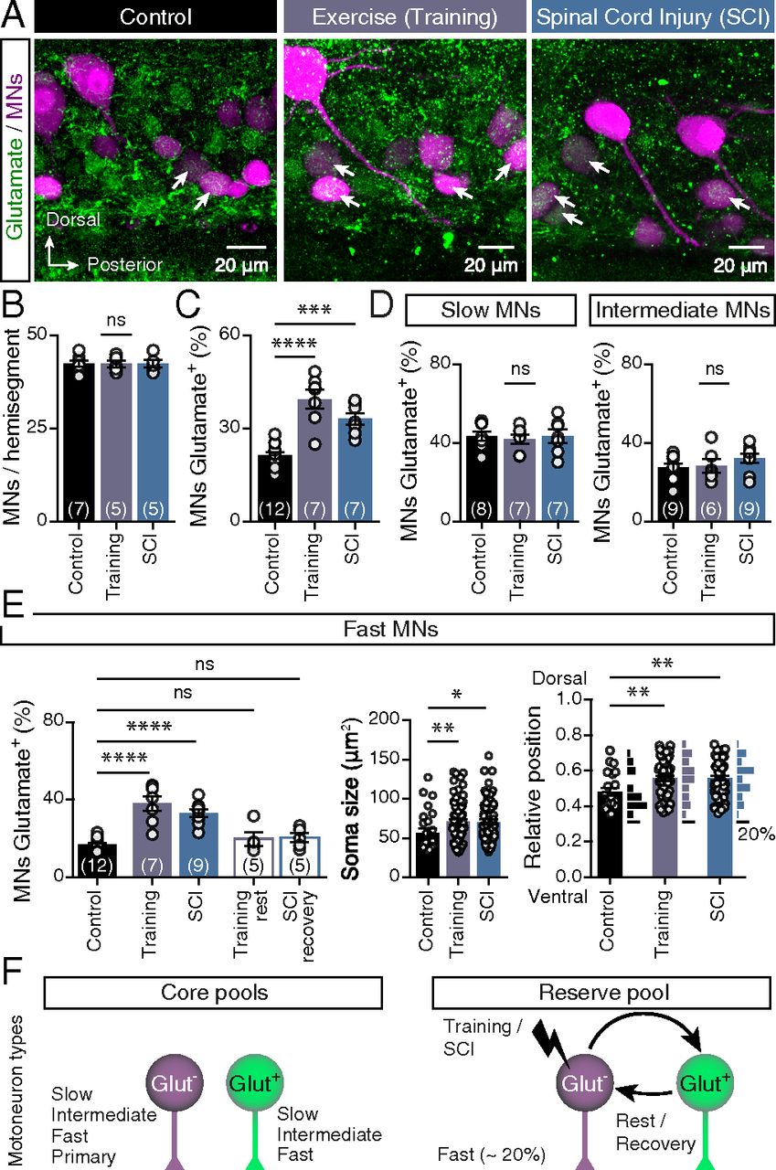
Respecification of motoneuron glutamatergic phenotype. (A) Representative whole-mount confocal images showing glutamatergic expression in axial motoneurons (MNs) of control animals, after swim training and after SCI. (B) Quantification of numbers of axial motoneurons. (C) Changes in percentages of axial motoneurons that coexpressed glutamate after swim training or SCI (P < 0.0001, one-way ANOVA). (D) The percentage of slow and intermediate motoneurons that expressed glutamate remained unchanged following these perturbations. (E) The percentage of glutamate+ fast motoneurons increased after training or SCI (P < 0.0001, one-way ANOVA). Quantification of soma sizes and locations of the glutamate+ fast motoneurons suggests that the larger motoneurons (P = 0.0099, one-way ANOVA) located in the dorsal part (P = 0.0089, one-way ANOVA) of the motor column retain ability to respecify their neurotransmitter phenotype. (F) Schematic representation of the three distinct pools of motoneurons observed in adult zebrafish spinal cord. Approximately 20% of the fast motoneurons constitute the reserve pool and retain ability to respecify their neurotransmitter phenotype. Arrows indicate the double-labeled cells. Data are presented as mean ± SEM; *P < 0.05; **P < 0.01; ***P < 0.001; ****P < 0.0001; ns, not significant. For detailed statistics see SI Appendix, Table S1.