
|
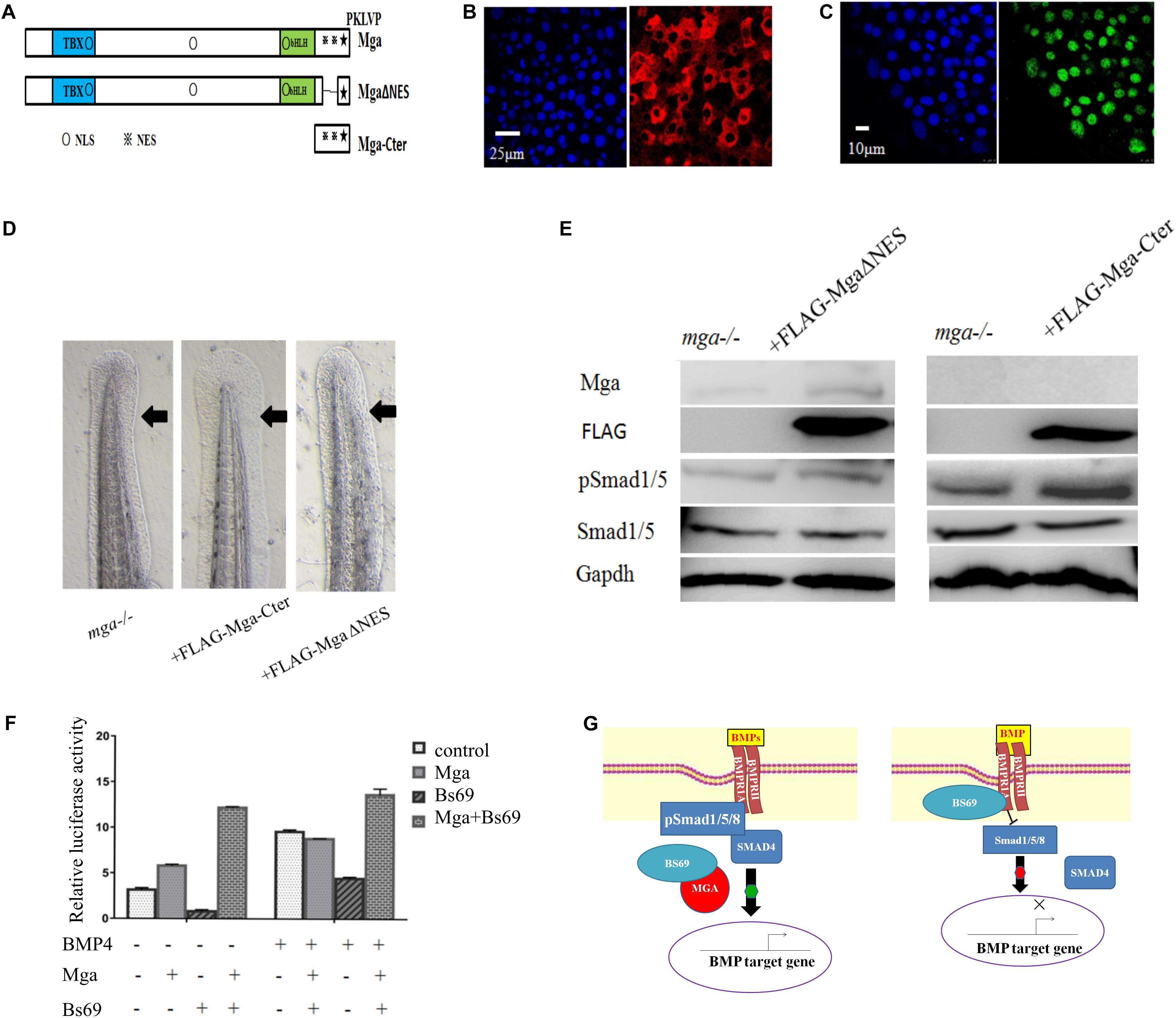
Fig. 6
Mga localized in the cytoplasm regulates Bmp signaling. (A) Diagram showing the location of putative NLS and ENS in Mga protein. ?: NLS, ?: NES. (B) IF showing the cytoplasmic localization of FLAG-Mga-Cter in 7 hpf embryos. (C) IF showing the nuclear localization of FLAG-Mga?NES in 7 hpf embryos. (D) FLAG-Mga-Cter but not FLAG-Mga?NES rescued the reduced ventral tailfin phenotype of mga mutant embryos at 2 dpf. (E) pSmad1/5 levels were increased in 7 hpf mga mutant embryos injected with 100 pg FLAG-Mga-Cter but not FLAG-Mga?NES. (F) The BRE-luc activity assay for Mga and Bs69 in C2C12 cells. (G) Cartoon model of how Mga regulates Bmp signaling. Left: Mga localized in the cytoplasm associates with Bs69 which allow Bmpr1a to phosphorylate and activate Smad1/5/8. The pSmad1/5/8 form complex with Smad4 and translocate into nuclei to regulate Bmp target gene expression. Right: In the absence of Mga, Bs69 associates with Bmpr1a and interferes with its phosphorylation and activation of Smad1/5/8 which caused reduced Bmp signaling. All experiments were performed in technical triplicate and are representative of multiple experiments.