Fig. 5
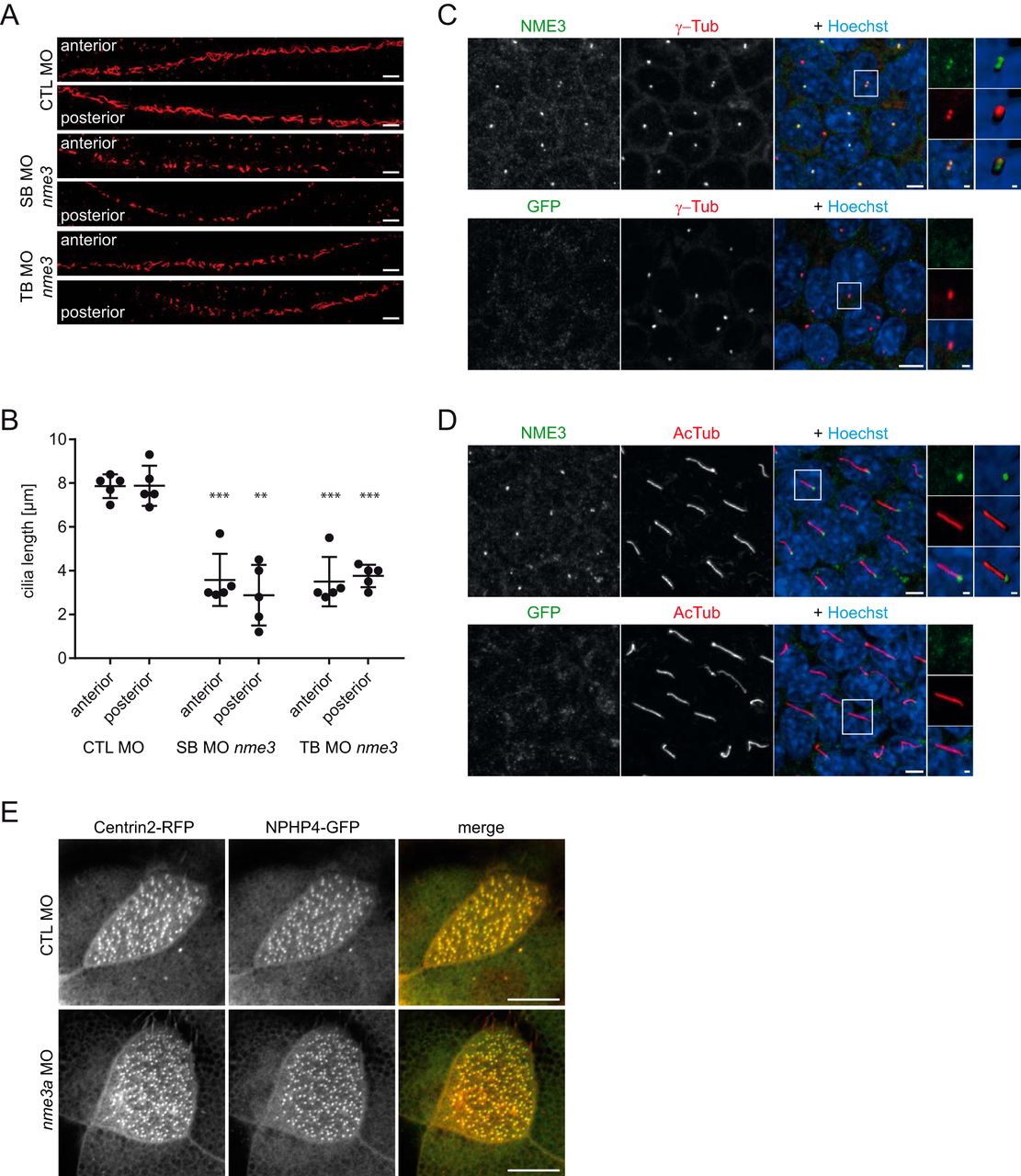
Nme3 is important for ciliogenesis in zebrafish and localizes to the ciliary basal body of mIMCD3 cells. A, confocal images of the anterior and posterior segments of the ciliated pronephric tubules of 24-hpf control (CTL) and nme3-deficient zebrafish embryos. Cilia were visualized by acetylated tubulin staining. Scale bars, 10 μm. B, quantification of the ciliary length in pronephric tubules. In contrast to the controls, ciliary length was affected by knockdown of nme3 (**, p = 0.0063; ***, p < 0.001; t test, error bars represent S.D.). C, confocal images of 6-day-starved mIMCD3 cells stained with antibodies against γ-tubulin (γ-Tub) (centrosomal marker; red), NME3 (green), and Hoechst (DNA; blue). NME3 colocalizes with γ-tubulin at the centrioles of centrosomes. D, costaining of cilia by acetylated tubulin (AcTub) (ciliary axoneme; red) identifies NME3 at the basal body of primary cilia. Immunofluorescence staining with an anti-GFP antibody serves as a control and results in the absence of centrosome and basal body labeling (bottom row). Scale bars, 5 μm. Magnifications and three-dimensional reconstructions are shown in white boxes. E, NPHP4–GFP and Centrin–RFP were coexpressed in epidermal multiciliated cells of stage 30 Xenopus embryos and detected by confocal microscopy. NPHP4 colocalization with the basal body marker Centrin was not altered by injection of nme3a morpholino in at least 25 cells observed in three experiments. Scale bars, 1 (C and D) and 10 μm (E).

