
Image
|
Figure Caption
Fig. S4
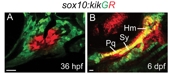
Fate mapping of the Fox-C expression domain
Images show the first two arches at 36 hpf (A) and the resultant skeleton at 6 dpf (B). sox10:kikGR+ arch CNCs (green) were photoconverted to red fluorescence with UV light using the ROI function on a Zeiss LSM800 confocal microscope. Photoconversion in a similar domain to where foxc1a and foxd1 are expressed resulted in labeling of the palatoquadrate (Pq), symplectic (Sy), and hyosymplectic (Hm) cartilages. Scale bars = 25 ?m.
Figure Data
Acknowledgments
This image is the copyrighted work of the attributed author or publisher, and
ZFIN has permission only to display this image to its users.
Additional permissions should be obtained from the applicable author or publisher of the image.
Full text @ Development