Fig. S3
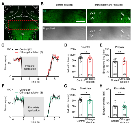
Effects of off-target ablation on intravenous general anesthesia, Related to Figure 2.
(A) Confocal image showing GFP expression in the brain of a 5-dpf Tg(HuC:GFP) larva. The dotted outlines indicate the borders of major brain regions. E, eye; HB, hindbrain; OT, optic tectum. Scale: 50 ?m.
(B) Enlarged images of the area outlined by a dashed red square in (A), showing that, after off-target ablation of neurons which were dorsal-lateral to the LC area at both the left and right hindbrain, GFP signals in the ablation sites disappeared (top, white arrows) and bulb-like structures appeared (bottom, white arrows). Scale: 50 ?m.
(C) Spontaneous swimming distance of control (black) and off-target ablated larvae (red) before, during and after the treatment of 30 ?M propofol. Each data point represents the total swimming distance for 5 min. Larvae at 6 dpf were used.
(D and E) Induction (D) and emergence time (E) of anesthesia induced by 30 ?M propofol for control (black) and off-target ablated larvae (red). Each symbol represents the data from a single larva.
(F) Spontaneous swimming distance of control (black) and off-target ablated larvae (greed) before, during and after the treatment of 30 ?M etomidate. Each data point represents the total swimming distance for 5 min. Larvae at 6 dpf were used.
(G and H) Induction (G) and emergence time (H) of anesthesia induced by 30 ?M etomidate for control (black) and off-target ablated larvae (green). Each symbol represents the data from a single larva. The number in the brackets indicates the number of larvae examined. The unpaired student?s t-test was performed for statistical analysis. n.s., not significant. Data are represented as mean ± SEM.

