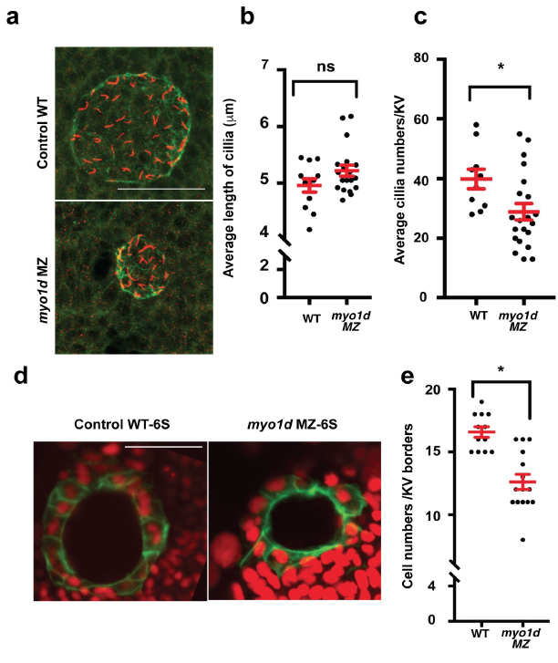
Fig. S4
Ciliogenesis appeared unaffected in zebrafish KV
a. Kupffer?s vesicle at 8 S embryos stained for aPKC (green) and acetylated tubulin (red). Cilia appeared crowded in myo1d MZ embryos compared to controls.
b. Average cilia length of KV were not significantly affected (n=31).
c. Average cilia numbers of KV were significantly less (n=31).
d. Tg(dusp6:GFP-MA) embryos injected with H2BmCherry mRNA showing KV epithelial cell morphology. In this experiment, number of nuclei bordering KV epithelial cell borders were counted.
e. Cell numbers lining KV borders at 6S significantly affected in myo1d MZ mutants. (n=28).

