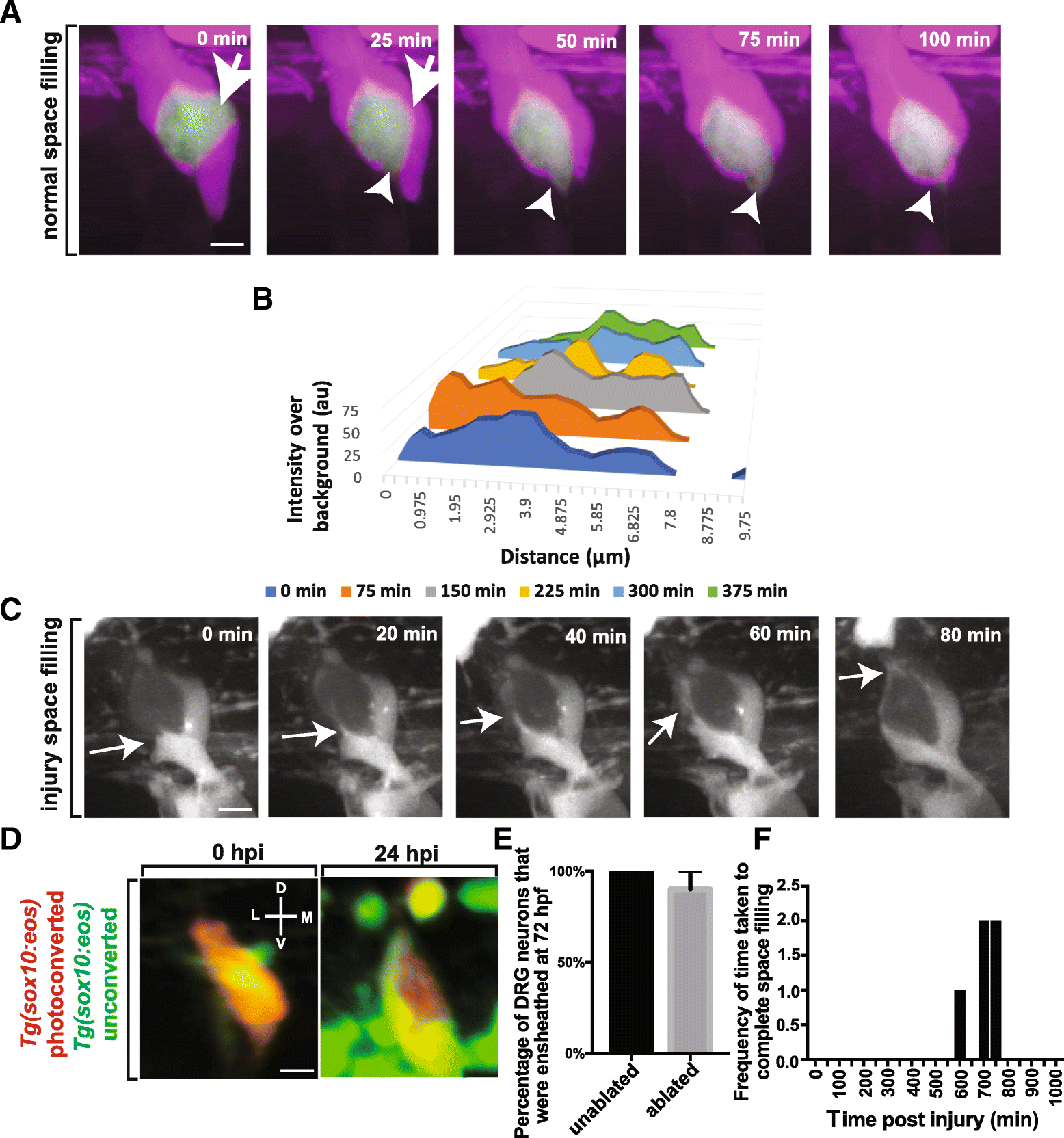
Fig. 4
Cells ensheathing DRG neuron cell somas exhibit space filling throughout life. (a). Confocal z-projection images from a 24-h timelapse starting at 72 hpf of a Tg(sox10:eos); Tg(ngn1:gfp) with photoconverted ensheathing cells. White arrowhead and arrows denotes areas of neuronal soma that are re-ensheathed. (b). Intensity profiles transecting two approaching ensheathing processes every 30 min. (c). Confocal z-projection images from a 24-h timelapse starting at 48 hpf of a Tg(sox10:eos) zebrafish with a laser ablation of an ensheathing cell. White arrows denote the migrating ensheathing process. (d). Confocal 3D images of a Tg(sox10:eos) DRG with a photoconverted neuron and an ablated ensheathing cell at 0 and 24 hpi. D denotes dorsal, M denotes medial, V denotes ventral, and L denotes lateral. (e). Percent of DRG neurons that are ensheathed 24 hpi (n?=?14 unablated DRG, n?=?10 ablated DRG). (f). Histogram of the time after ablation that re-ensheathment of the neuron soma is completed (n?=?5 DRG). Scale bar is 10 ?m (a, c, d)

