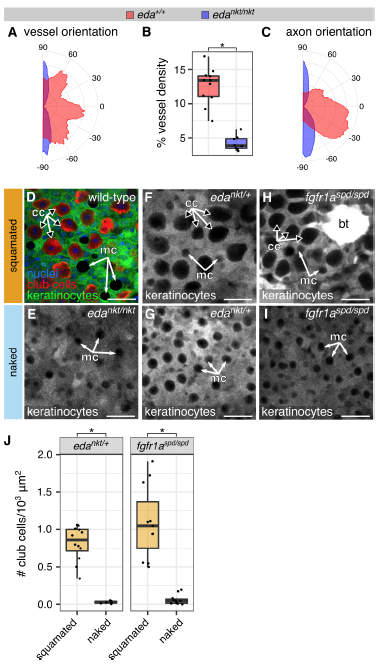
Fig. S7
Eda and Fgfr1a are required for cutaneous neurovascular polarization and maturation, related to Figure 7
(A-C) Orientation and density analysis of skin vasculature and innervation from projections through intact skin of animals expressing endothelial [Tg(fli1a:EGFP)] (A,B) and axonal [Tg(p2rx3a>mCherry)] markers (C). *, p<0.01, Wilcoxon rank sum test. n=8-11 skin regions of size 0.77-1.5 mm2 from n=3-5 fish analyzed per genotype. (D) Single z-plane through the epidermis of an isolated scale immunostained as indicated. Note that club cells (cc) and mucus secreting cells (mc) do not express the keratinocyte marker Tg(krt4:EGFP) and can be distinguished by their characteristic shapes and sizes. (E-I) Single z-planes through the epidermis of intact fish of the indicated genotypes. Note the lack of club cells in the “naked” (non-squamated) skin regions. Tg(krt4:EGFP) is highly expressed in breeding tubercles (bt), male-specific keratin-rich structures visible in panel H. cc, club cells; mc, mucus secreting cells. (J) Quantification of club cell density based on the pattern of Tg(krt4:EGFP) expression in naked and squamated skin of fish of the indicated genotypes. *, p<0.01, Wilcoxon rank sum test. n=6-12 skin regions of size 0.1-1.2 mm2 from n=4-6 fish analyzed per condition. Transgenes: (D-I) keratinocytes [Tg(krt4:EGFP)]. Staining: (D) club cells (CS-56) and nuclei (DAPI). Scale bars, 25 μm.
Reprinted from Developmental Cell, 46(3), Rasmussen, J.P., Vo, N.T., Sagasti, A., Fish Scales Dictate the Pattern of Adult Skin Innervation and Vascularization, 344-359.e4, Copyright (2018) with permission from Elsevier. Full text @ Dev. Cell

