Fig. 4
Fig. 4
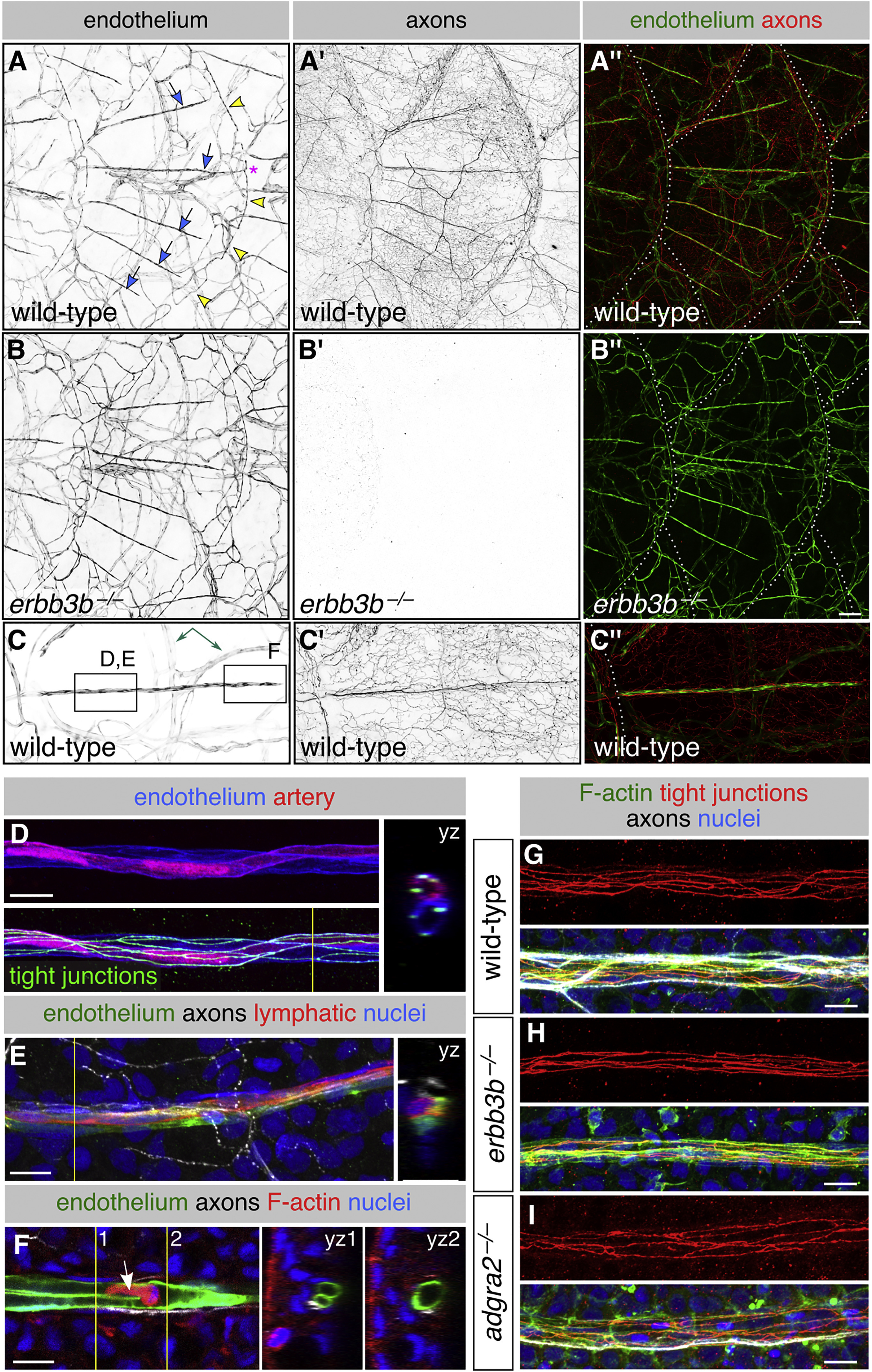
Scales Vascularize Independently of Sensory Axons
(A?C) Representative lateral views of the trunk from wild-type and erbb3b?/? adults. Arrows, superficial scale capillaries. Arrowheads, microvasculature associated with scale margin. Asterisk, interconnection of superficial capillary with scale margin vasculature. Dashed lines, scale margins. Double-headed arrow, example of dermal vasculature not associated with sensory nerves. Boxes in (C) indicate areas of magnification for (D)?(F).
(D and E) Magnification of scale capillaries. Note the two intertwined vessels in (D).
(F) Single z section through the tip of a scale capillary. Orthogonal views show that vessel lumens merge near posterior tip to form a U shape. Arrow, cell within the vessel lumen. Yellow lines in (D)?(F), planes of orthogonal sections.
(G?I) Representative scale capillaries immunostained for the indicated markers from adult wild-type, erbb3b?/? and adgra2?/? fish.
Transgenes: endothelium (Tg(fli1a:EGFP)); (A?C) axons (Tg(p2rx3a>mCherry)); (D) artery (Tg(-0.8flt1:RFP)) (Bussmann et al., 2010); lymphatic (TgBAC(prox1a:KALTA4,4xUAS-ADV.E1b:TagRFP)) (van Impel et al., 2014). Staining: (D) tight junctions (anti-Tjp1); (E) axons (acTubulin) and nuclei (DAPI); (F) F-actin (phalloidin), axons (acTubulin), and nuclei (DAPI); (G?I) F-actin (phalloidin), tight junctions (anti-Tjp1), axons (acTubulin), and nuclei (DAPI). Scale bars, 100 ?m (A?C) and 10 ?m (D?I). See also Figure S4.
Reprinted from Developmental Cell, 46(3), Rasmussen, J.P., Vo, N.T., Sagasti, A., Fish Scales Dictate the Pattern of Adult Skin Innervation and Vascularization, 344-359.e4, Copyright (2018) with permission from Elsevier. Full text @ Dev. Cell

