
|
Fig. S1
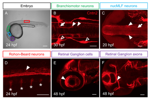
Cntn2 protein expression during embryonic development
(A) Lateral view of a 24 hpf embryo with boxes indicating the location of different neuronal cell types in B-F. Panels B-F show confocal projections of embryos labeled with anti-Cntn2 antibody (red). Panel B shows a dorsal view, and C and F show ventral views. Panels D and E show lateral views. All panels show anterior to the left. (B) A 30 hpf embryo showing Cntn2 expression in FBM neuron cell bodies in r6 and r7 (arrowheads), and their axons (arrow). Open arrowheads mark sensory ganglia. (C) A 20 hpf embryo showing Cntn2 expression in nucMLF cell bodies (arrowheads), and their axons (arrow). (D) A 24 hpf embryo showing Cntn2 expression in central axons (arrowhead) of Rohon-Beard (RB) neurons. Asterisks indicate axons of primary motor neurons exiting the spinal cord. (E, F) A 48 hpf embryo showing Cntn2 expression in nasal RGC (arrowhead, E) as well as RGC axons (arrowhead, F). Scale bars: A, 200 ?m, and B-F, 50 ?m.
Reprinted from Mechanisms of Development, 152, Gurung, S., Asante, E., Hummel, D., Williams, A., Feldman-Schultz, O., Halloran, M.C., Sittaramane, V., Chandrasekhar, A., Distinct roles for the cell adhesion molecule Contactin2 in the development and function of neural circuits in zebrafish, 1-12, Copyright (2018) with permission from Elsevier. Full text @ Mech. Dev.