
|
Fig. 3
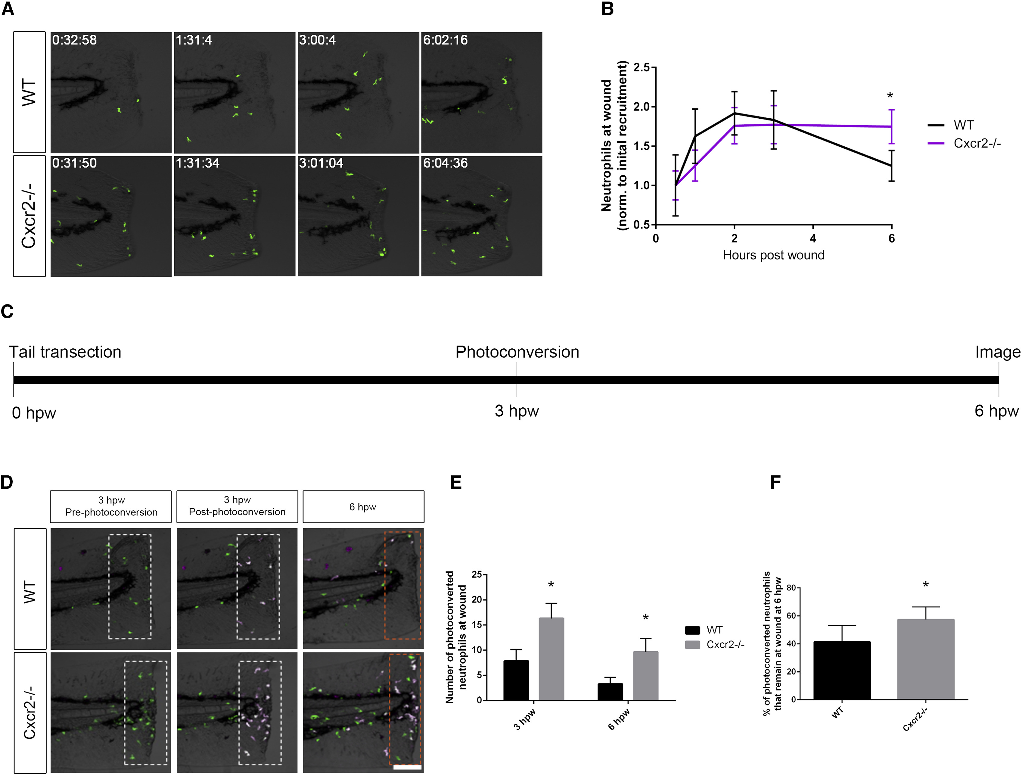
Neutrophil Reverse Migration Is Impaired in cxcr2 Mutants
(A) Time-lapse imaging of Tg(mpx:Dendra) WT and Cxcr2?/? tail fins after wounding.
(B) Quantification of average neutrophil presence at wound normalized to the average initial neutrophil recruitment to wound at 30 min post-wound (n = 11 WT and 14 mutant; one representative experiment). WT larvae exhibit neutrophil recruitment to wound peaking at 2 hpw with resolution back to 30 min post-wound levels by 6 hpw, whereas Cxcr2?/? larvae maintain high levels of neutrophil infiltration in the wound microenvironment at 6 hpw.
(C) Schematic of photoconversion of Cxcr2?/? neutrophils at wound. Tg(mpx:Dendra) Cxcr2?/? larvae were wounded at 3 dpf. Photoconversion of Dendra+ neutrophils within the wounded tail fin (white dotted outlines in D) was performed at 3 hpw, and neutrophil reverse migration was assessed at 6 hpw.
(D) Neutrophils pre- and post-photoconversion at 3 hpw and at 6 hpw were imaged in WT and Cxcr2?/?.
(E) Quantification of photoconverted neutrophils in the wound microenvironment (magenta cells in dotted boxes, D; n = 25 WT and 30 mutants). cxcr2?/? wounds contained a higher level of photoconverted cells at 3 and 6 hpw.
(F) Percent of photoconverted cells that remained at the wound at 6 hpw was calculated. Cxcr2?/? neutrophils remain at the wound at a higher frequency than WT neutrophils. ?p < 0.05.
Error bars represent SE.