Fig. S2
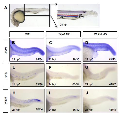
Rspo1, runx1, wnt16 levels in Rspo1 MO or Wnt16 MO animals. (A) Image of WT embryo at 24 hpf showing area of images taken and labeled with the location of the neural tube (NT, gray), dorsal aorta (DA, red line), posterior cardinal vein (PCV, blue line), and yolk sac (YS). Representative wild type (WT; B,E,H), Rspo1 MO (C,F,I), and Wnt16 MO (D,G,J) embryos processed by WISH for rspo1 (B-D), runx1 (E-G), and wnt16 (H-J) mRNA expression at 22 hpf or 24 hpf as indicated in each image. Lateral views of the trunk at 160x magnification. Numbers of embryos displaying the depicted phenotype are indicated.

