Fig. S5
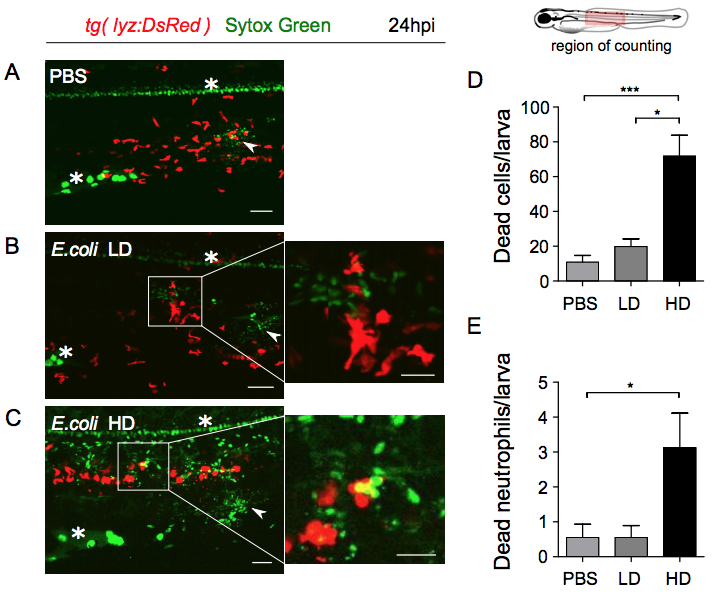
Notochord infection induces neutrophils death.
Tg(lyz:DsRed) larvae were either injected with PBS (A) or infected with low dose (LD) (B) or high dose (HD) (C) of E. coli in the notochord. Neutrophils were detected using DsRed (red) and dead cells using Sytox Green (green) at 24 hpi and trunk regions were imaged using Spinning Disk Confocal microscopy. Representative maximal projections of confocal montages show increased cell death, including dead neutrophils around the notochord in HD infection, comparing to LD and PBS injection. White stars show non-specific staining in the yolk extension and neurones of the spinal cord. Arrowheads show Sytox Green injection sites. White boxes in the left panels show the zoomed areas (right panels). Scale bars: 50 ?m for the left panels and 25 ?m for the right panels. (D) Number of Sytox Green positive cells and (E) Sytox Green positive neutrophils around the notochord in indicated conditions (mean number of cell/larva ± SEM, NPBS = 9, NLD = 9 and NHD = 8, from two independent experiments, Kruskal Wallis test with Dunn?s post-test, *p<0.05, ***p<0.001).

