
|
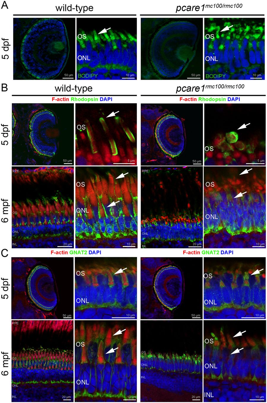
Fig. 3
Pcare1rmc100/rmc100 zebrafish show aberrant photoreceptor morphology. (A) Analysis of the morphology of the pcare1rmc100/rmc100 zebrafish larval retina (5 dpf) using boron-dipyrromethene (BODIPY) revealed disorganization of photoreceptor outer segments as compared to those of strain- and age-matched wild-type larvae (arrows). (B) Siblings of adult zebrafish without (wild-type) or containing (pcare1rmc100/rmc100) the 29 bp deletion in pcare1 were sectioned and stained with antibodies against F-actin (red), Rhodopsin (green) or, in (C) GNAT2 (green). Arrows indicate normal outer segments in control fish and dysmorphic outer segments in mutant fish. Nuclei were stained with DAPI.