
|
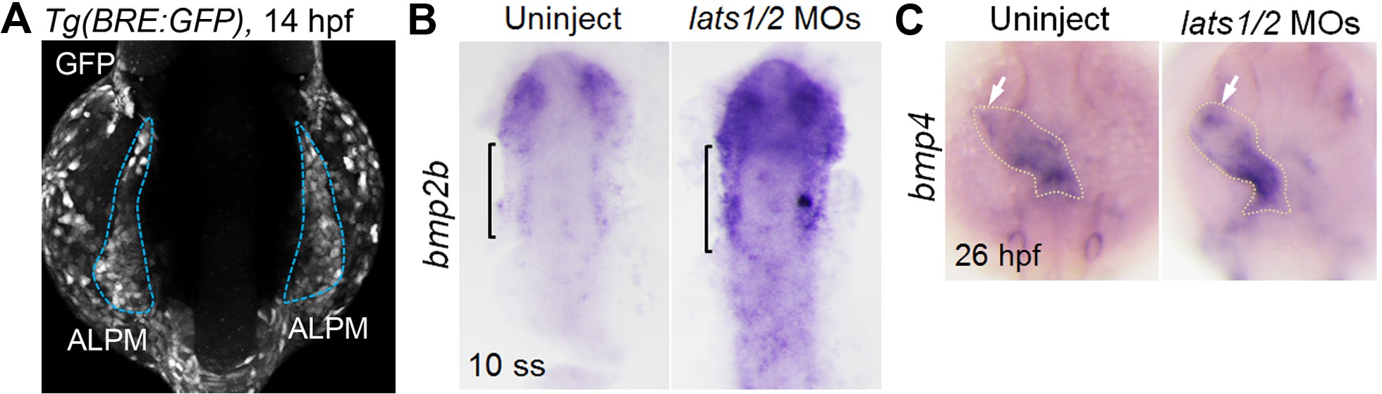
Fig. 6-S1
Depletion of Lats1/2 leads to an increase in bmps expression.
(A) Confocal 3D-stack image of the Tg(BRE:GFP) embryo at 14 hpf (n = 6). Blue broken lines indicate the GFP-positive ALPM. (B, C) WISH analyses at 10 ss (B) and 26 hpf (C) of uninjected control embryos (left panels, n = 15 to 22) and embryos injected with lats1/2 MOs (right panels, n = 19 to 23) made using antisense probes for bmp2b (B) and bmp4 (C). (B) Square brackets indicate the bmp2b-positive ALPM. Note that expression of bmp2b is increased in the lats1/2 morphants. (C) Arrows indicate the bmp4-positive cells in the venous pole. Broken lines indicate the cardiac tube. The confocal 3D-stack image and WISH images are a set of representative images from at least three independent experiments.