
|
Fig. 5
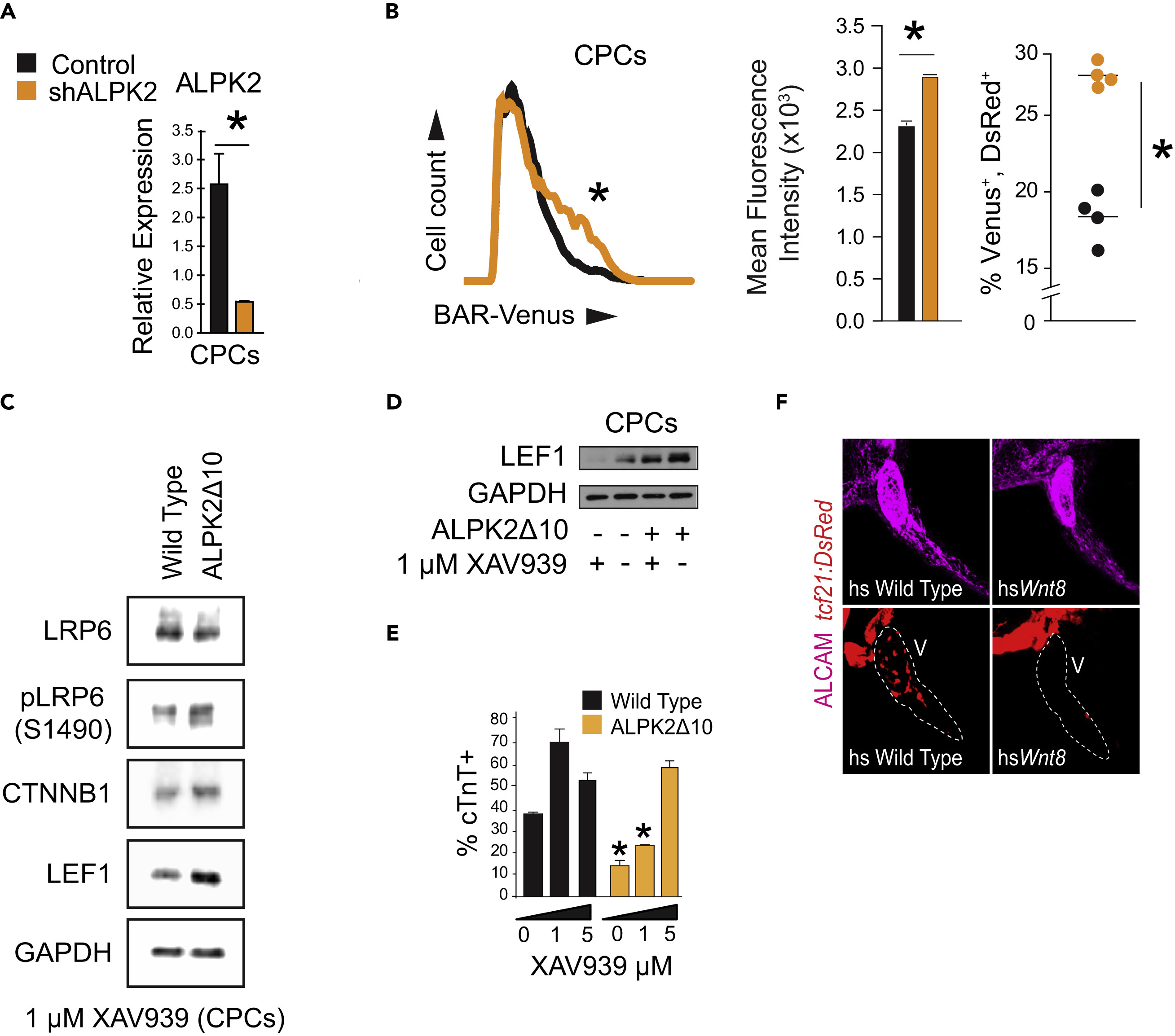
ALPK2 Inhibits WNT/?-Catenin Signaling to Promote Cardiogenesis
(A) Transcripts abundance of ALPK2 in control and ALPK2 shRNA-transfected cardiac progenitor cells (CPCs) derived from human embryonic stem cells (hESCs).
(B) Flow cytometric plot and graphs quantifying venus expression driven by ?-catenin activated reporter (BAR) in live CPCs following control or ALPK2 shRNA transfection as hESCs.
(C) Western blot protein expression analysis of WNT/?-catenin signaling modulators in wild-type and CRISPR/Cas9-mediated ALPK2 mutant hESC-derived CPCs (ALPK2?10) treated with a small molecule direct tankyrase inhibitor XAV939 (1 ?M).
(D) Western blotting for the WNT/?-catenin signaling target LEF1 in wild-type and ALPK2?10 cells with or without the addition of XAV939 (0 or 1 ?M).
(E) TNNT2 flow cytometry quantification of day 14 cardiomyocytes derived from wild-type and ALPK2?10 hESCs treated with XAV939 (0, 1, 5 ?M).
(F) Representative images of heat shocked (hs) wild-type and hsWnt8b transgenic zebrafish carrying a transgene for the epicardium (tcf21:DsRed) at 96 hpf. Fish were counterstained with an activated leukocyte cell adhesion molecule (ALCAM; magenta).
N = 3?5 biological replicates, and data are displayed as mean � SEM. * = p ? 0.05. See also Figure S4.