
|
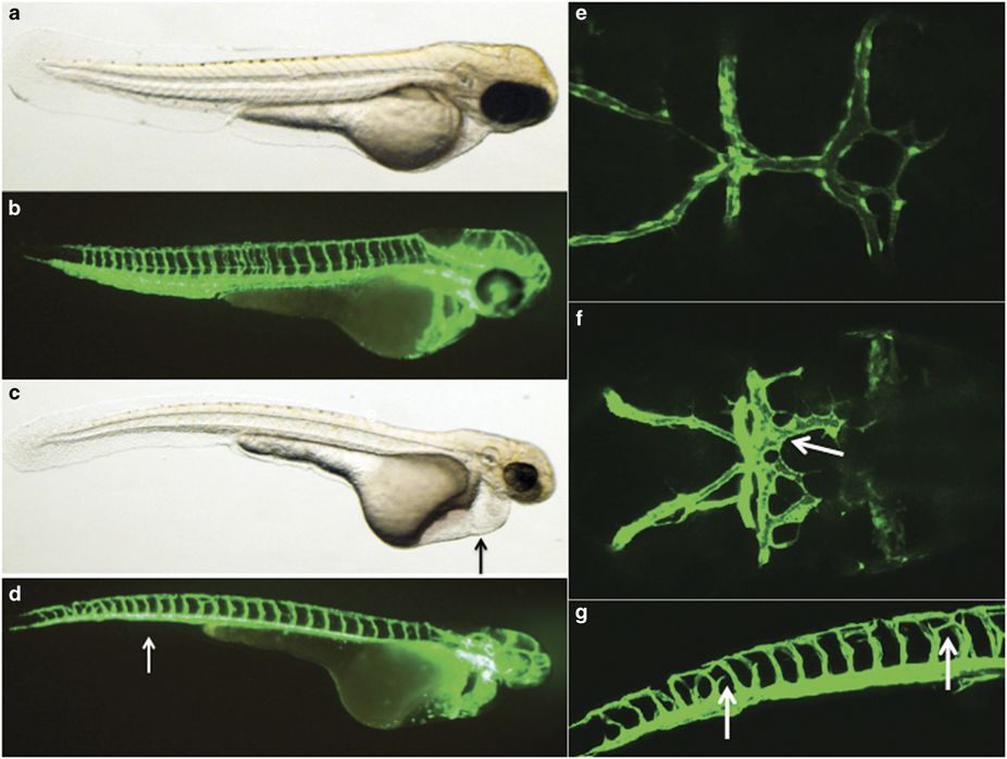
Fig. 4
Zebrafish modeling of the SMAD9 mutation. Zebrafish morphants resulting from an injection of a translation blocking morpholino were created and imaged with a confocal microscope at 68?72?h post fertilization. Uninjected (a) light and (b) confocal lateral imaging of Tg(kdrl:eGFP) embryos was performed; the embryos were oriented laterally, cranial-right. Comparative images of SMAD9 knockdown (c, d) were typified by smaller heads, smaller eyes and thin trunks. The embryos also demonstrated cardiac edema (c, arrow) and had a near-absence of the caudal vein plexus (d, arrow). Cranial circulation was also imaged in uninjected (e) and morphants (f) with embryos oriented dorsal, cranial-right. Numerous abnormal arteriovenous connections between the dorsal longitudinal vein, mesencephalic vein and metencephalic artery were present (arrow). Further evidence of abnormal artery?vein specification during development was observed in the intersegmental vessels, where ectopic branches (g, arrow) were observed above the myoseptum.