
|
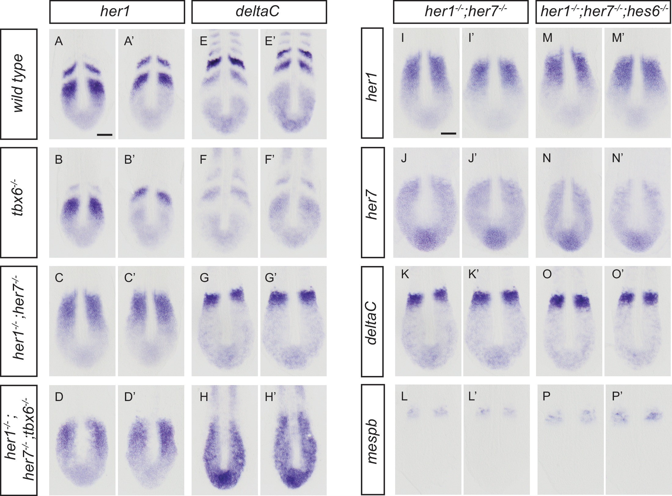
Fig. 1-S2
Disruption of the segmentation clock in tbx6, her1;her7, her1;her7;tbx6 and her1;her7;hes6 mutants.
(A-H') In situ hybridization for segmentation clock markers her1 and deltaC in tbx6?/?, her1?/?;her7?/?and her1?/?;her7?/?;tbx6?/?. her1 (B and B?) and deltaC (F and F?) oscillate in the posterior PSM of tbx6?/?. her1 (C and C?) and deltaC (G and G?) do not oscillate in her1?/?;her7?/?. (D and D?, H and H?) her1 and deltaC do not oscillate and their expression is restricted to the posterior PSM in her1?/?;her7?/?;tbx6?/?. (I-P') Comparison of clock markers her1, her7 and deltaC and segmental output marker mespb between her1?/?;her7?/? and her1?/?;her7?/?;hes6?/?. her1, her7 and deltaC expression domains are indistinguishable in her1?/?;her7?/? (I and I?, J and J?, K and K?) and her1?/?;her7?/?;hes6?/? (M and M?, N and N?, O and O?). mespb expression in the anterior PSM is equivalently disordered in both mutants (L and L?, P and P?). Embryos are 13.5 hpf (10-somite stage). Scale bar in A applies to A-H?. Scale bar in I applies to I-P?. Scale bars are 100 �m.