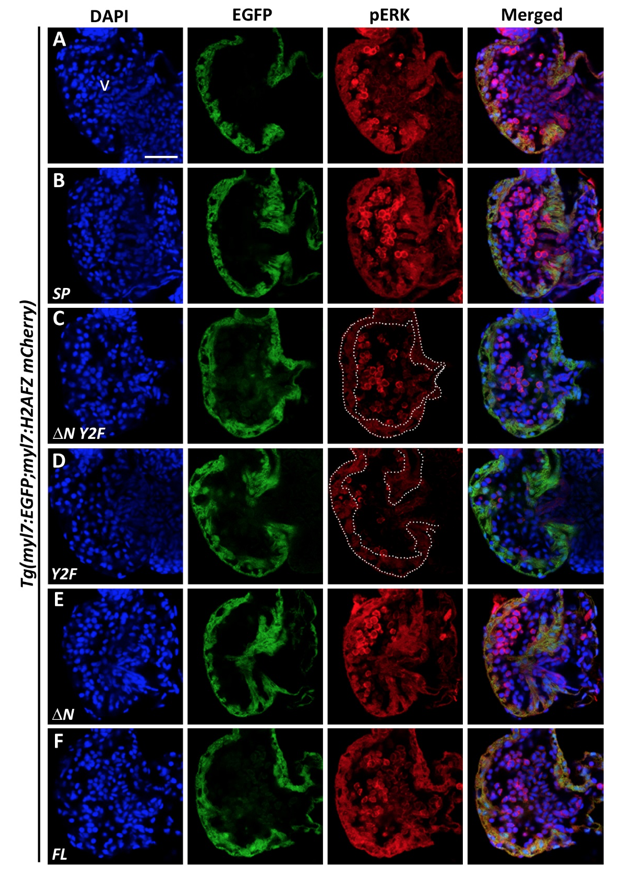
Fig. S6
Endogenous expression of pERK was nearly abolished in heart ventricles of ?N Y2F or Y2F VCAP1X2 mRNA-overexpressing Tg(myl7:EGFP; myl7:H2AFZ mCherry) embryos. Decreased expression level of pERK was detected in heart ventricles of Tg(myl7:EGFP; myl7:H2AFZ mCherry) embryos that overexpressed ?N Y2F (C) or Y2F VCAP1X2 (D) mRNA compared to un-injected (A) or transgenic embryos that overexpressed SP (B), ?N (E) or FL (F) VCAP1X2 mRNA at 96 hpf (n = 15 per condition, N = 3). Dashed lines indicate the ventricular myocardium region. Erythrocytes in the ventricular chamber show non-specific staining from secondary antibody. Nuclei were labeled by DAPI staining. v, ventricle. Scale bar, 30 ?m.

