
|
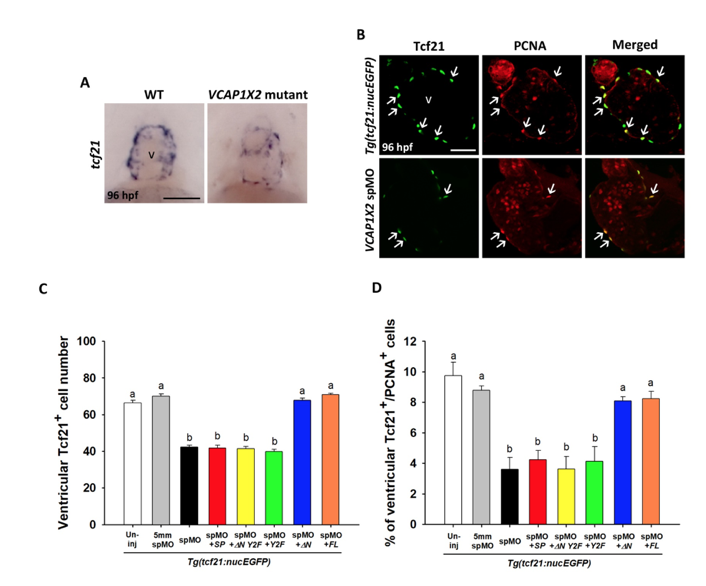
Fig. S8
Epicardium formation was affected in VCAP1X2 mutants and morphants, but could be rescued by co-injection of △N or full-length (FL) VCAP1X2 mRNA. (A) Epicardium defects were detected by tcf21 staining in the ventricle of VCAP1X2 mutant, but not wild-type (WT) heart at 96 hpf (n = 15 per condition, N = 3). Scale bar, 50 μm. v, ventricle. (B) Tcf21+ epicardial cell number was reduced with coincident decreases in PCNA+ cell number in VCAP1X2 spMO-injected Tg(tcf21:nucEGFP) transgenic embryos compared to un-injected transgenic embryos at 96 hpf. Arrows indicate Tcf21+/PCNA+ epicardial cells. Scale bar, 25 μm. (C) Quantification of Tcf21+ epicardial cell number in ventricles of Tg(tcf21:nucEGFP) transgenic embryos was performed following different treatments (n = 20 per condition, N = 3). (D) The percentage of PCNA+ proliferative epicardial cells in ventricles of Tg(tcf21:nucEGFP) transgenic embryos following different treatments was compared (n = 20 per condition, N = 3). Error bars indicate standard error. Quantitative data were analyzed by ANOVA with Bonferroni multiple comparisons. Treatments that are not statistically different (α = 0.05) from each other are labeled with the same letter.