
|
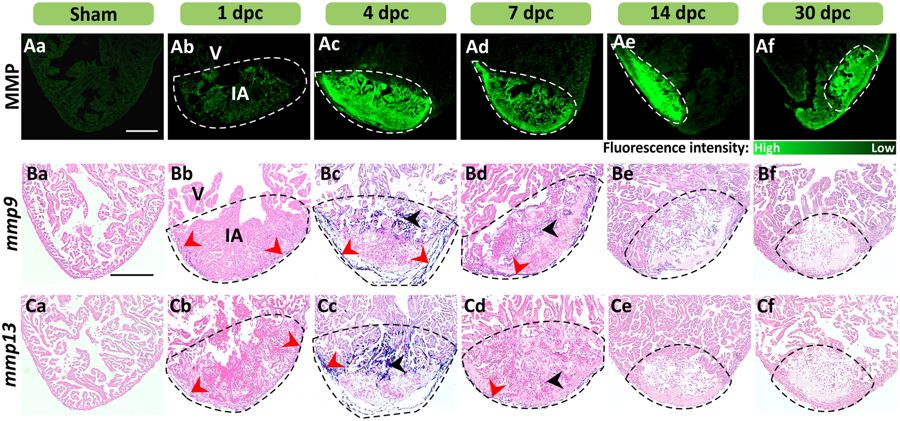
Fig. 1
MMP activity and mmp9 and mmp13 expression are enhanced at sites of injury in the heart. (A) In situ zymography was conducted to show the activity of MMP (in green) in representative cryosections of (Aa) intact hearts and in (Ab?Af) injured hearts at 1?30 dpc. (B,C) In situ hybridization was conducted to show the expression of (B) mmp9, and (C) mmp13 (blue labeling), in representative paraffin sections of (Ba, Ca) intact and (Bb?Bf, Cb?Cf) cryoinjured ventricles at 1?30 dpc. The sections were counterstained with Fast Red (pink labeling) to label all the cells. The red and black arrowheads indicate regions of mmp expression in the epicardium and myocardium, respectively. In each panel, the regions bounded by the dashed lines indicate the injured area (IA) of the ventricle (V). Scale bars: 200?�m.