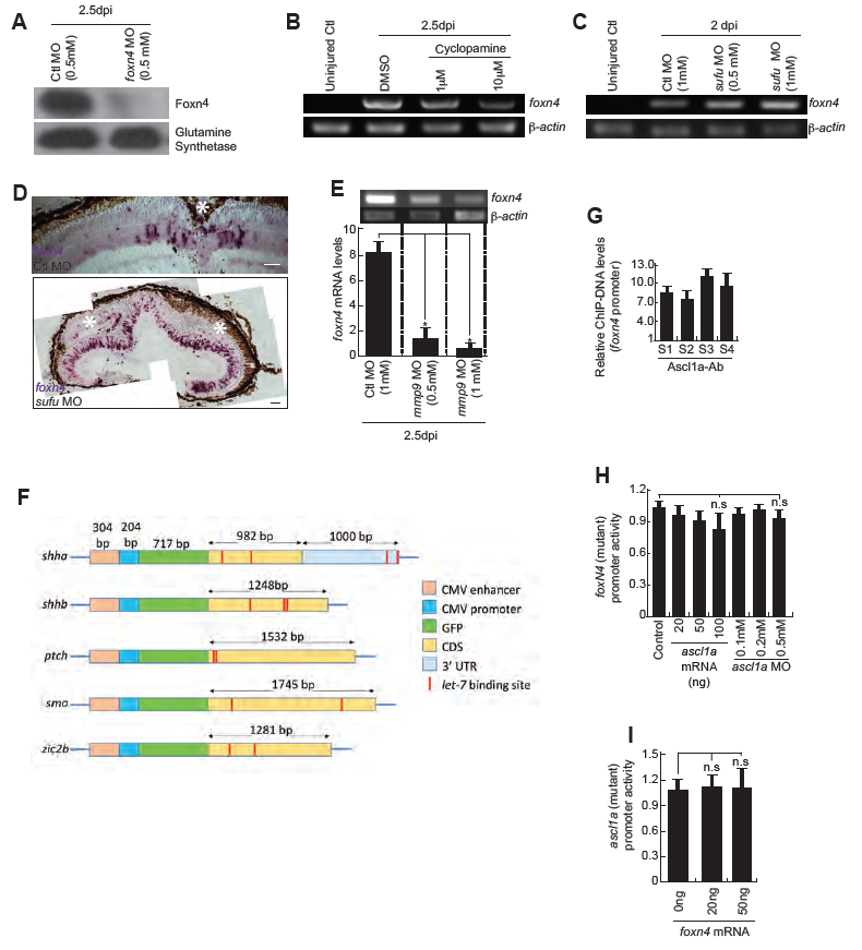
Fig. S5
Expression of foxn4 in retina at various conditions (A) Western blot analysis of Foxn4 in foxn4-MO electroporated retina, at 2.5dpi. (B) RT-PCR analysis of foxn4 in uninjured control, 2.5dpi DMSO-treated, and 2.5dpi cyclopamine-treated whole retina. (C) RT-PCR analysis of foxn4 from sufu MOelectroporated retina compared with control MO, at 2dpi. (D) BF microscopy images of foxn4 mRNA ISH in retinal sections electroporated with control and sufu MOs at 4dpi. (E) RT-PCR (upper) and qPCR (lower) analysis of foxn4 in control MO, and mmp9 MO electroporated in 2.5dpi retina. *P<0.001 in E, and error bars are SD. (F) Schematic representation of DNA constructs used in transfection experiments for examining the impact of let-7 microRNA on various genes. (G)qPCR assay revealed the relative abundance of ChIP DNA fragments of foxn4 promoter obtained by Ascl1a antibody which are normalized to control uninjured retina. (H) Luciferase assay revealed that mutated Ascl1a-BS on foxn4 promoter had little effect on positive or negative regulation by ascl1a mRNA or MO respectively. (I) Luciferase assay revealed that mutated Foxn4-BS on ascl1a promoter had little effect on positive regulation by foxn4 mRNA. Scale bars, 10 ?m (D).

