Fig. S1
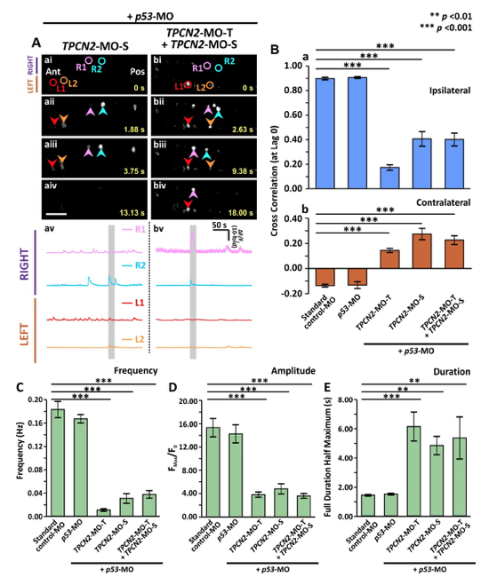
Effect of MO-based knockdown of TPC2 (via injection of either TPC2-MO-S or TPC2-MO-S and TPC2-MO-T) on the spontaneous Ca2+ activity of CaPs in SAIGFF213A;UAS:GCaMP7a double-transgenic embryos at ~24 hpf. SAIGFF213A;UAS:GCaMP7a embryos were injected with: (Aa) TPCN2-MO-S with p53-MO; or (Ab) TPCN2-MO-T and TPC2-MO-S with p53-MO. (Aai-Abiv) Time-lapse fluorescence images showing the changes in GCaMP7a fluorescence in the CaPs at different time intervals in the two treatment groups. The embryos are in a dorsal orientation and regions of interest (ROIs) on two selected CaP cell bodies on the left (L) and right (R) sides of the spinal cord are shown. The arrowheads indicate GCaMP7a signals in the CaPs. Ant. and Pos. are anterior and posterior, respectively. Scale bar is 50 Ám. (Aav,Abv) Line graphs to show the ?F/F0 against time (over a period of ~300 sec) in the ROIs of the representative embryos shown in (Aai-Abiv), respectively. The time period that corresponds to the Ca2+ signaling events shown in the time-lapse fluorescence images (Aai-Abiv) is denoted by a grey vertical bar in (Aav-Abvi). (B) Bar graphs to show the mean ▒ SEM cross correlation function at zero lag of the (Ba) ipsilateral, and (Bb) contralateral CaP Ca2+ activity. (C-E) Bar graphs to show the mean ▒ SEM (C) frequency, (D) amplitude and (E) duration of the Ca2+ spikes in the CaPs of the double-transgenic embryos in the various treatment groups. Data were obtained from n=24-63 cells from 4-7 embryos, except for the duration data, which were n=6 cells from 3 embryos. p<0.01 (**) and p<0.001 (***), and NS indicates no significant difference between the data.
Reprinted from Developmental Biology, 438(1), Kelu, J.J., Webb, S.E., Galione, A., Miller, A.L., TPC2-mediated Ca2+ signaling is required for the establishment of synchronized activity in developing zebrafish primary motor neurons., 57-68, Copyright (2018) with permission from Elsevier. Full text @ Dev. Biol.

