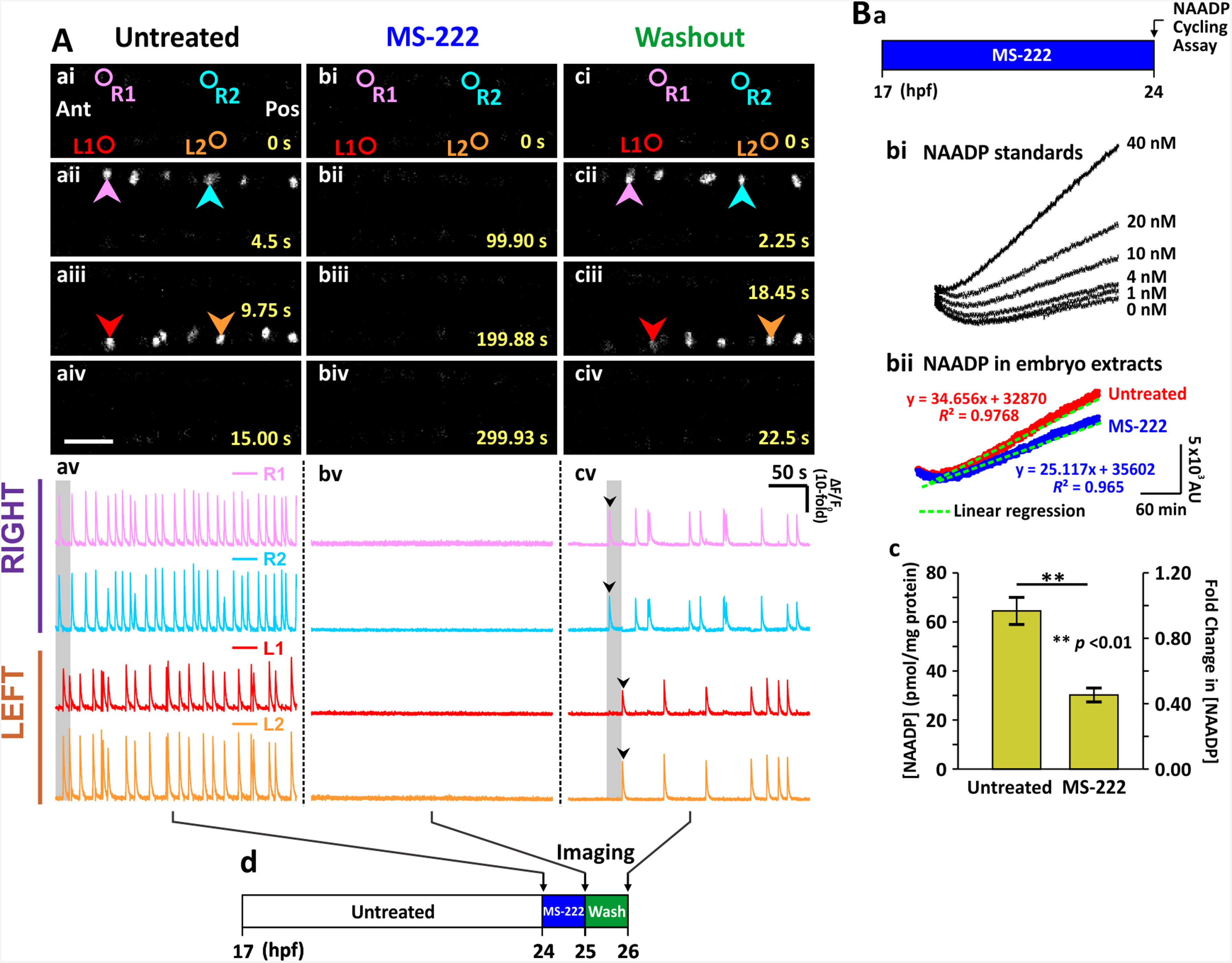
Fig. 7
Effect of inhibiting the action potentials on the spontaneous Ca2+ activity of the CaP in SAIGFF213A;UAS:GCaMP7a embryos, and on the production of NAADP by AB wild-type embryos, at ~24 hpf. SAIGFF213A;UAS:GCaMP7a embryos were (Aa) imaged at ~24 hpf. (Ab) MS-222 was then applied for 1?h and the embryos were imaged again (i.e., at ~ 25 hpf). (Ac) Subsequently, the MS-222-treated embryos were washed with Danieau?s solution for 1?h, after which they were imaged one more time (i.e., at ~26 hpf). (Aai-Aciv) Time-lapse fluorescence images, and (Aav-Acv) line graphs as described in Fig. 1. The data presented in panels Aa-Ac were collected from the same representative embryo. (Ad) Schematic to show the timing of the MS-222 treatment and washout experiments. (B) The NAADP cycling assay was used to detect the endogenous level of NAADP in embryos at ~24 hpf. (Ba) Schematic to show the timing of the MS-222 treatment. (Bb) Line graphs show the change in the fluorescence intensity of resofurin over time (covering a period of ~ 270?min) such that (Bbi) shows NAADP standards of 0?40?nM, and (Bbii) shows NAADP measured in samples prepared from intact embryos at ~24 hpf in the absence/ presence of MS-222. These plots were then fitted into linear regression models, and the slope (m) of fluorescence increase was determined by y?=?mx+c. (Bc) Bar graph showing the normalized [NAADP], and the corresponding fold-change of [NAADP] in the samples prepared from embryos±?MS-222 (n?=?9 from 3 independent assays). p?<?0.01 (**).
Reprinted from Developmental Biology, 438(1), Kelu, J.J., Webb, S.E., Galione, A., Miller, A.L., TPC2-mediated Ca2+ signaling is required for the establishment of synchronized activity in developing zebrafish primary motor neurons., 57-68, Copyright (2018) with permission from Elsevier. Full text @ Dev. Biol.

