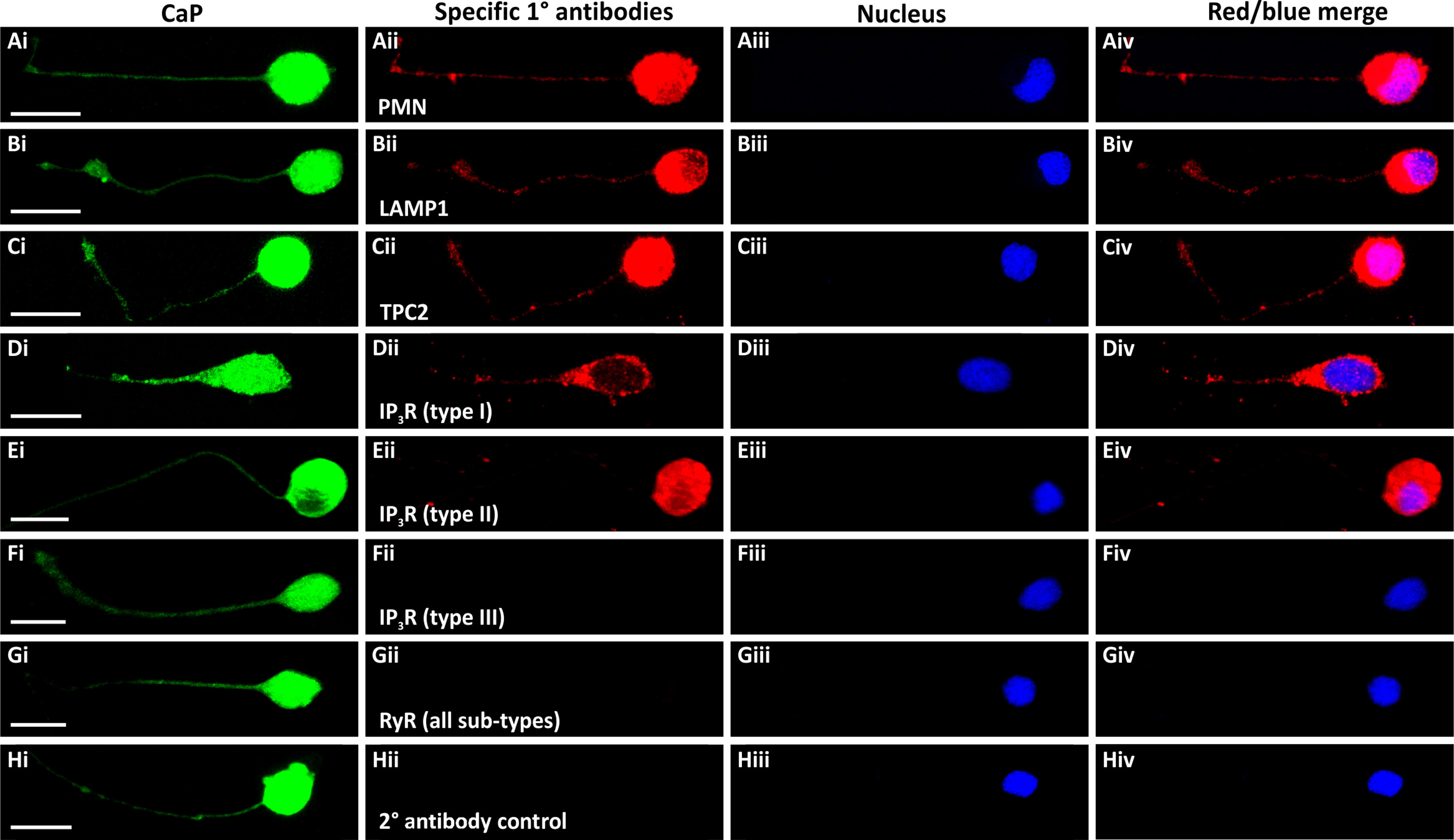
Fig. 6
Expression and localization of LAMP1, TPC2, IP3Rs and RyRs in presumptive CaPs isolated from SAIGFF213A;UAS:GFP embryos and cultured for 24?h. The trunk of these embryos, which (Ai-Hi) express GFP in the CaPs, was dissected at ~18 hpf. The cells were dissociated and cultured for 24?h and then they were immunolabelled with antibodies for: (Aii) PMNs, (Bii) lysosomal-associated membrane protein 1 (LAMP1), (Cii) TPC2, (Dii-Fii) IP3R types I-III, respectively, or (Gii) RyR (all sub-types). Panel (Hii) shows a secondary antibody control. (Aiii-Hiii) The cells were co-stained with DAPI to label the nuclei. Each panel is a series of optical sections that have been projected as a single confocal image, and they show: (Ai-Hi) GFP in the presumptive CaPs (in green); (Aii-Hii) the localization of the various proteins immunolabelled (in red); (Aiii-Hiii) the DAPI-labeled nuclei (in blue); and (Aiv-Hiv) images showing the red and blue channels when merged. Scale bars, 10?Ám. (For interpretation of the references to color in this figure legend, the reader is referred to the web version of this article)
Reprinted from Developmental Biology, 438(1), Kelu, J.J., Webb, S.E., Galione, A., Miller, A.L., TPC2-mediated Ca2+ signaling is required for the establishment of synchronized activity in developing zebrafish primary motor neurons., 57-68, Copyright (2018) with permission from Elsevier. Full text @ Dev. Biol.

