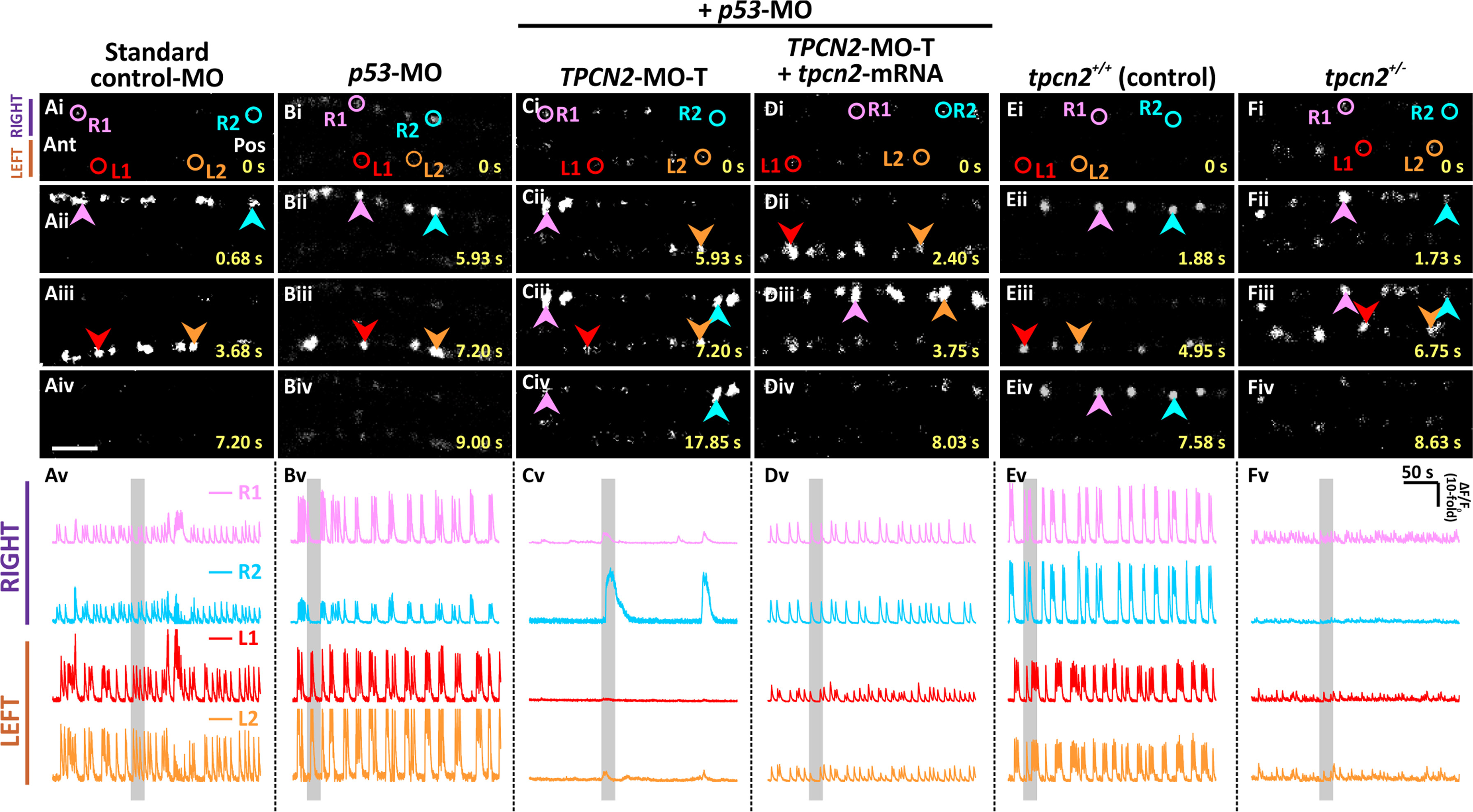
Fig. 1
Effect of TPC2-knockdown, heterozygous-knockout, and mRNA rescue on the spontaneous Ca2+ activity of the CaPs in SAIGFF213A;UAS:GCaMP7a embryos at ~24 hpf. SAIGFF213A;UAS:GCaMP7a embryos were injected with: (A) Standard control-MO; (B) p53-MO; (C) TPCN2-MO-T with p53-MO; or (D) TPCN2-MO-T with p53-MO and the mutant tpcn2-mRNA. In addition, (E) untreated SAIGFF213A;UAS:GCaMP7a fish (termed tpcn2+/+ controls) were imaged and (F) they were crossed with homozygous tpcn2dhkz1a mutants to generate double-transgenic heterozygous mutants (tpcn2+/-). (Ai-Fiv) Time-lapse fluorescence images showing the changes in GCaMP7a fluorescence in the CaPs at different time intervals in the various treatment groups. The embryos are in a dorsal orientation and regions of interest (ROIs) on two (of the three) selected CaP cell bodies on the left (L) and right (R) sides of the spinal cord are shown. The arrowheads indicate GCaMP7a signals in the CaPs. Ant. and Pos. are anterior and posterior, respectively. Scale bar, 50?Ám. (Av-Fv) Line graphs showing the ?F/F0 against time (over a period of ~ 300?s) in the ROIs of the representative embryos shown in (Ai-Fiv), respectively. The time period that corresponds to the Ca2+ signaling events shown in the time-lapse fluorescence images (Ai-Fiv) is denoted by a grey vertical bar in (Av-Fv). Also see Movies S1-S3, which illustrate the distinct right/left alternation of Ca2+ transients in the controls and how this is disrupted when TPC2-mediated Ca2+ release was inhibited.
Reprinted from Developmental Biology, 438(1), Kelu, J.J., Webb, S.E., Galione, A., Miller, A.L., TPC2-mediated Ca2+ signaling is required for the establishment of synchronized activity in developing zebrafish primary motor neurons., 57-68, Copyright (2018) with permission from Elsevier. Full text @ Dev. Biol.

