
|
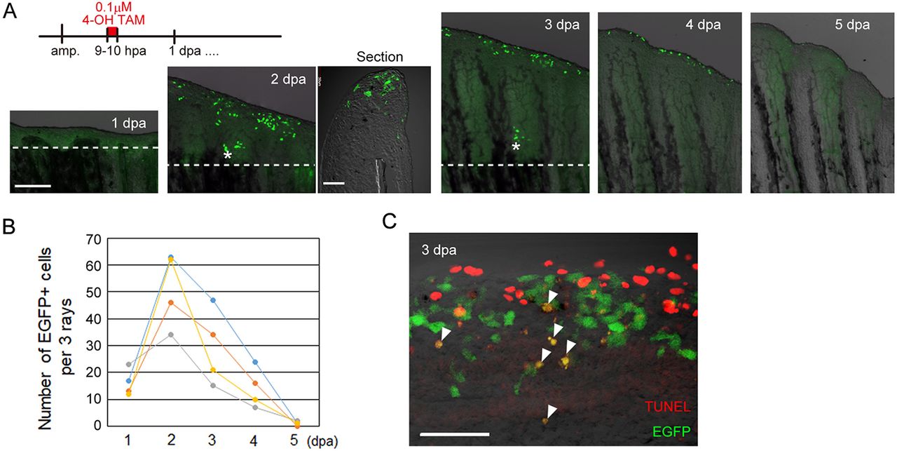
Fig. 3
Early RE cells are disposed of by apoptosis by 5?dpa during zebrafish fin regeneration. (A) Tracking of early RE cells that were pulse-labelled at 9-10?dpa with 0.1?�M 4-OH TAM. The labelled cells moved distally and disappeared by 5?dpa. The longitudinal section of the 2?dpa fin shows that the EGFP+ cells localize to the tip of the wound epidermis. The dotted lines indicate the amputation planes. Scale bars: 200?�m (fins) and 50?�m (section). (B) Quantification of the number of EGFP+ cells in A. EGFP+ cells in three adjacent rays were counted (n=4 zebrafish). (C) Detection of the TUNEL+ cells (red) and EGFP+ cells derived from the early RE cells that were labelled by Cre-loxP recombination at 9-10?hpa. The arrowheads show the TUNEL+ and EGFP+ cells. Scale bar: 50?�m.