
|
Fig. 7
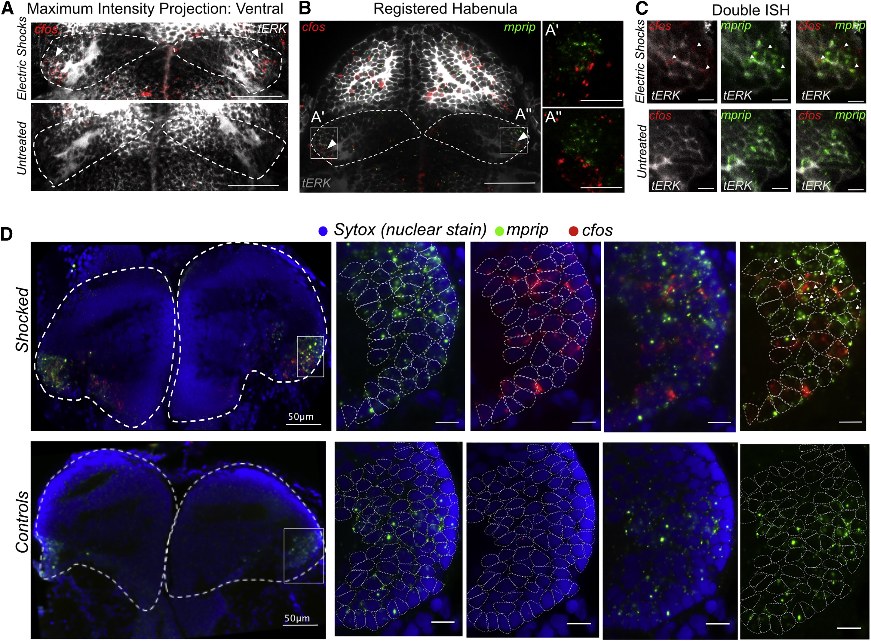
Noxious Electric Shocks Activate a Sub-population of Neurons in the Ventro-lateral Habenula Labeled by mprip
(A) ISH analysis of cfos expression in the habenula 30 min after exposure to electric shocks. (Scale bars represent 50 ?m.)
(B) Registration of cfos signals to habenular molecular atlas reveals co-regionalization with the mprip+ ventrolateral population. (Scale bars represent 50 ?m).
(C) Double ISH for c-fos and mprip (marker for ventrolateral neuronal type) showing a co-localization of cfos+ and mprip+ domains in the larval habenula in response to electric shocks. (Scale bars represent 10 ?m).
(D) Double ISH of cfos and mprip showing the conservation of electric shocks-induced cfos responses in mprip+ ventro-lateral neuronal type in the adult habenula. Nuclei borders are demarcated in the zoomed in panels on the right using dotted circles. (Scale bar represents 10 ?m unless otherwise stated).
See also Figure S7 and Movie S2.