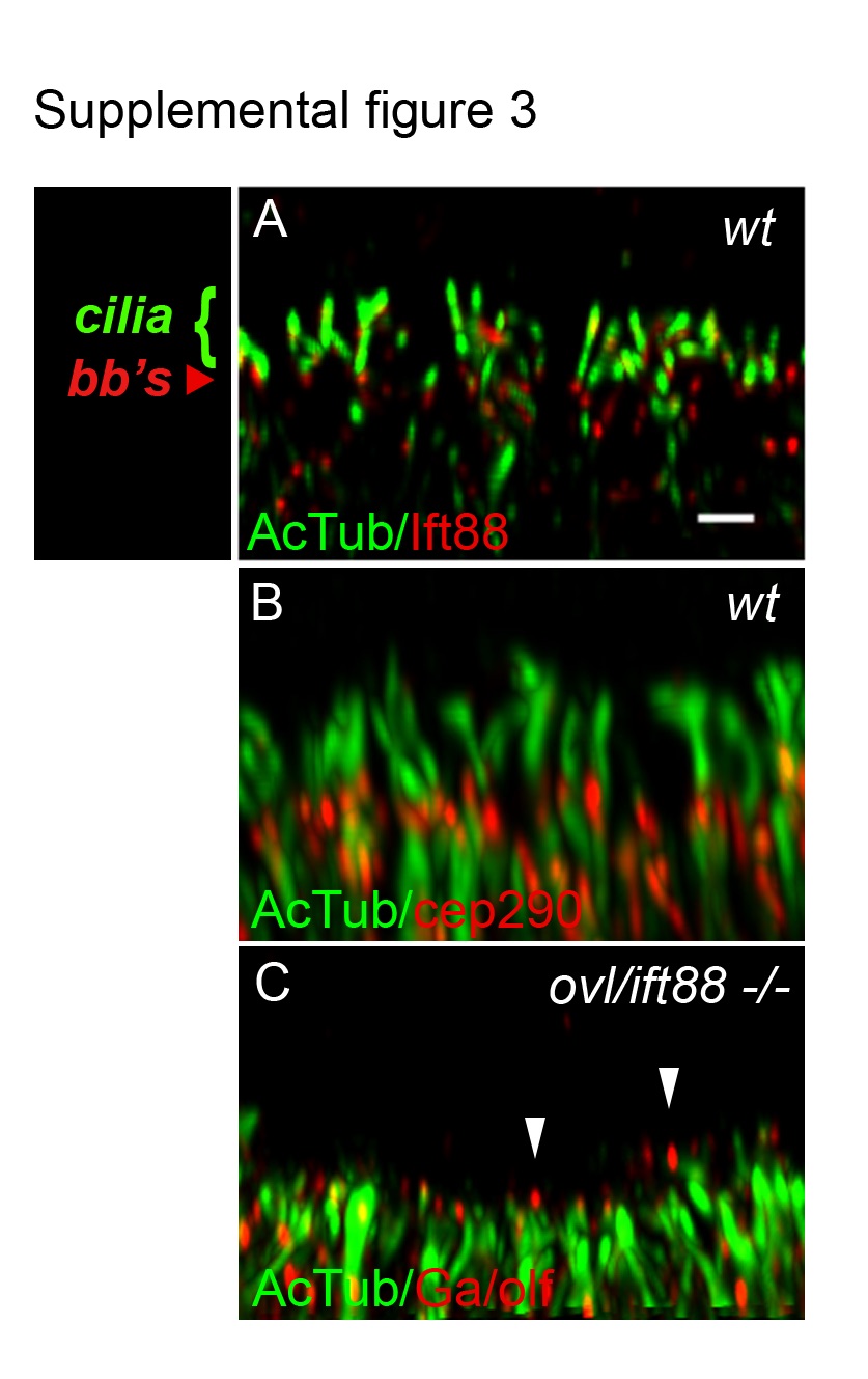
Fig. S3 Olfactory sensory cilia deficit in oval/ift88 -/- mutants. (A) Olfactory sensory cilia in the center of a wild type olfactory placode stained with anti-Ift88 (red) and anti-acetylated tubulin (green; cilia) and imaged in confocal Z-stacks. Ift88 immunoreactivity was strongest in basal bodies (bb's). Scale bar in (A) equals 1 Ám. (B) Wild type olfactory placode stained with anti-acetylated tubulin (green; cilia and neuronal cell body processes) and anti-cep290 (red; basal bodies). (C) oval mutant olfactory placode stained with anti-G /olf (red) and anti-acetylated tubulin (green) shows cilia loss with some short, residual G /olf-positive axonemes (arrowheads). All panels are set at equivalent scale and represent a 3Ám thick maximum intensity projection of the center of the olfactory placode.
Image
Figure Caption
Acknowledgments
This image is the copyrighted work of the attributed author or publisher, and
ZFIN has permission only to display this image to its users.
Additional permissions should be obtained from the applicable author or publisher of the image.
Full text @ Cilia

