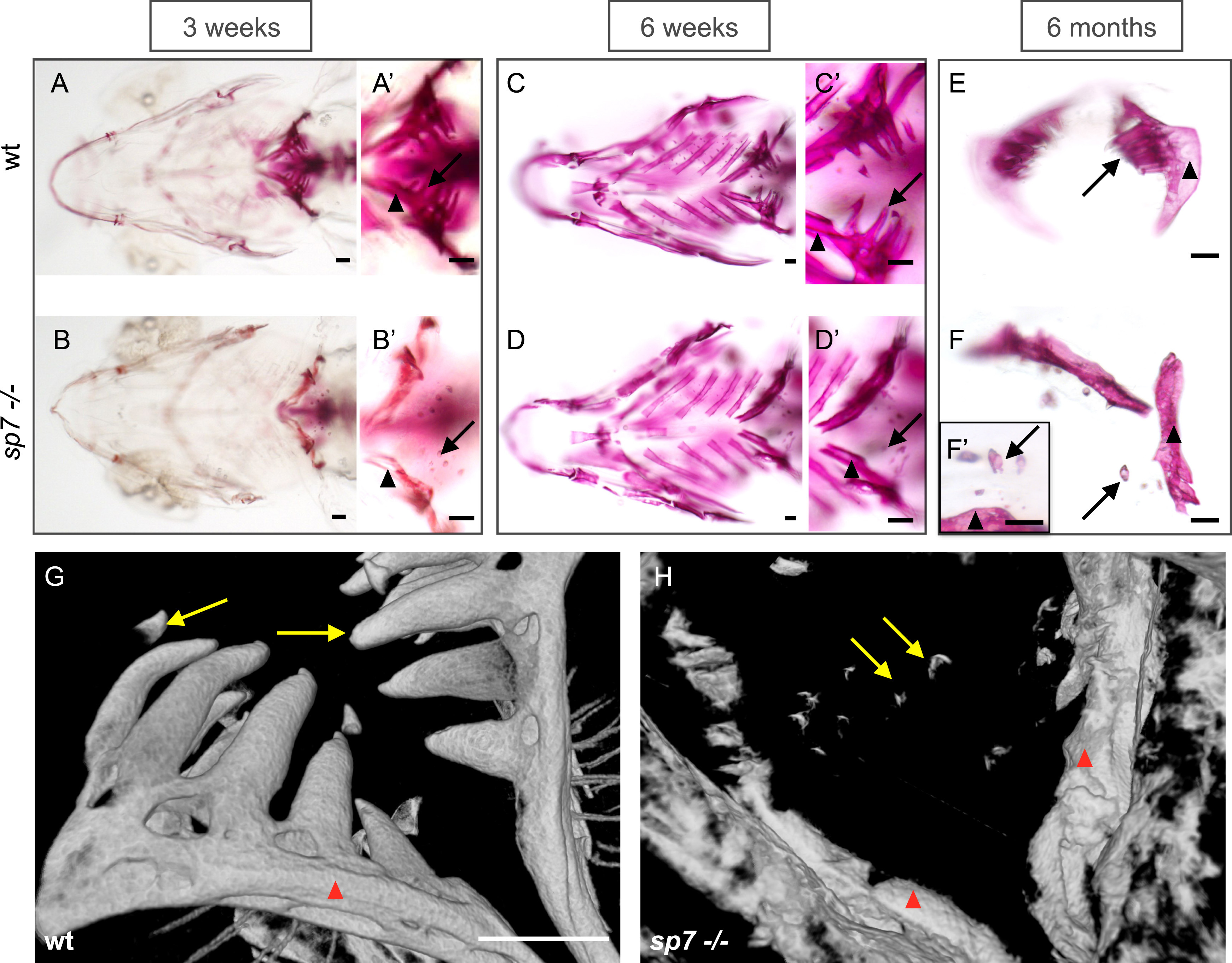
Fig. 1
Defective tooth formation and lack of attachment insp7mutants observed throughout life. Alizarin Red S staining to show mineralization of the zebrafish pharyngeal teeth and fifth ceratobranchial (pharyngeal jaw) (A-F). A, A?, B, B?) In comparison to the wt, the fifth ceratobranchial (arrowheads) is poorly mineralized in mutants at 3 weeks post-fertilization, with only the extreme tip of the teeth stained (arrows). C, C?, D, D?) At 6 weeks, mineralization of the fifth ceratobranchial has increased in the mutants, but teeth remain as before. E, F, F?) sp7 mutant pharyngeal bone displays a more narrow and thinner morphology, and teeth do not have a mineralized attachment to the bone. G, H) Volume rendering pictures of micro-computed tomography (ÁCT) of zebrafish (>1.5 year) to show pharyngeal bones (arrowheads) and teeth (arrows) in wt (G) and sp7 mutant (H). Note the shape of the pharyngeal bones in the sp7 mutant and the absence of any bone of attachment. Scale bars represent 100?Ám.
Reprinted from Developmental Biology, 435(2), Kague, E., Witten, P.E., Soenens, M., Campos, C.L., Lubiana, T., Fisher, S., Hammond, C., Brown, K.R., Passos-Bueno, M.R., Huysseune, A., Zebrafish sp7 mutants show tooth cycling independent of attachment, eruption and poor differentiation of teeth, 176-184, Copyright (2018) with permission from Elsevier. Full text @ Dev. Biol.

