Fig. 2-S2
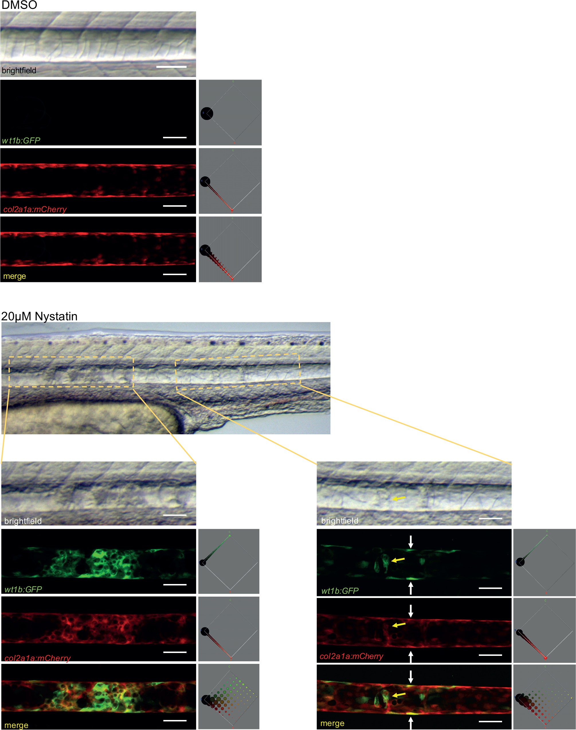
Nystatin treatment leads to upregulation of wt1b:gfp expression in notochord sheath cells.
48hpf Tg(wt1b:gfp; R2-col2a1a:mCherry);nacre-/- zebrafish embryos were treated with either 20 ÁM nystatin or 0.4% DMSO for 24 hr. The notochord structure of DMSO-treated embryos appeared normal (n = 15), while nystatin-treated embryos exhibited notochord lesions of varying severity along the notochord (n = 12/13). Confocal Z-stacks through the centre of the notochords show wt1b:gfp expression (green), R2-col2a1a:mCherry expression (red) and their overlap (merged). wt1b:gfp expression was observed at lesion sites, while no wt1b:gfp expression was observed in control animals. Colour histograms (Color-Inspector 3D ImageJ plugin) quantify pixel colour distribution, and show overlap between green and red expression in merged channel images. Experimental replicates = 3; n = 7?13 embryos/per treatment.

