Fig. 2
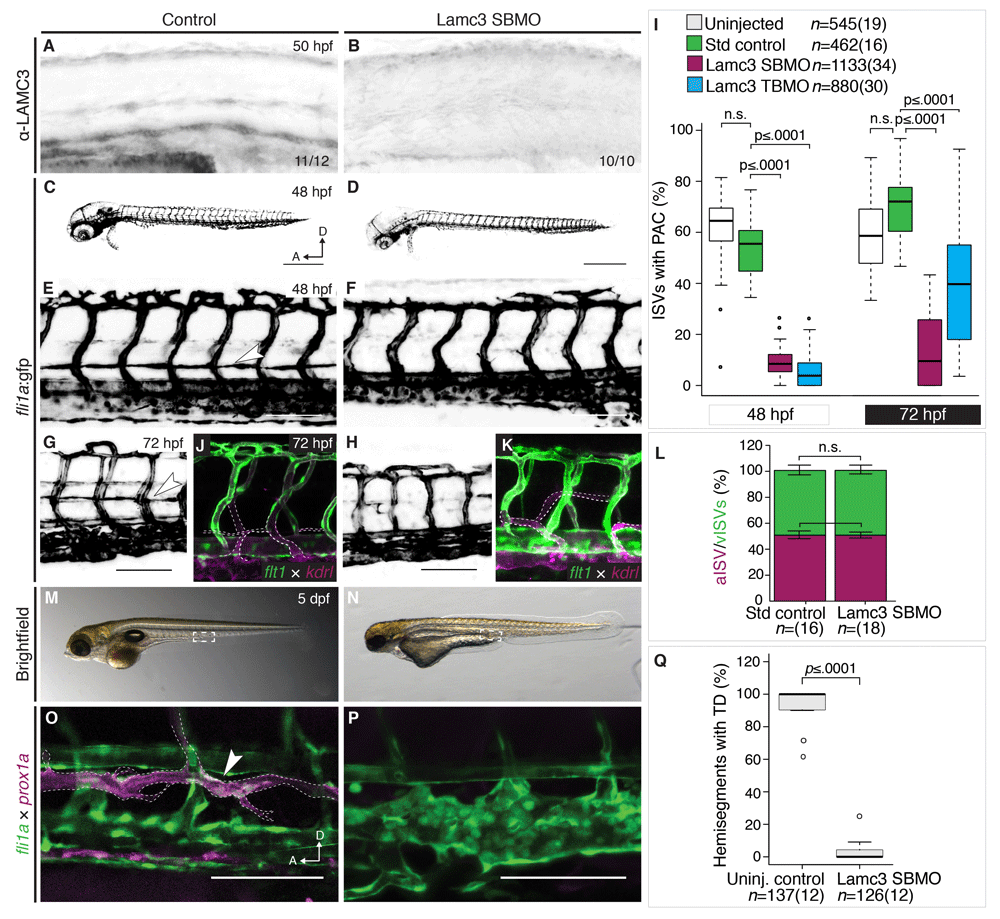
Lamc3 knockdown embryos do not develop the parachordal chain (PAC).
(A?B) Lateral view of the zebrafish trunk stained using anti-LAMC3 antibody shows reduction in Lamc3 protein at 50 hpf in knockdown embryos (E, n=10) compared to standard controls (D, n=11). (C?I) Vasculature of 48 hpf Tg(fli1a:egfp) embryos. (C) Embryos injected with standard control MO develop the PAC. (D) Lamc3 knockdown embryos do not develop the PAC. (E) An enlarged image of the trunk vasculature in control embryos the PAC is indicated by a white arrowhead. (F) An enlarged image of the trunk vasculature in Lamc3 SBMO injected embryos. (G) The parachordal chain in control embryos at 72 hpf (white arrowhead). (H) By 72 hpf Lamc3 embryos have still not developed a parachordal chain. (I) Quantification of all ISVs with a PAC in Tg(fli1a:egfp) uninjected (white, n=545), control (green, n=462), Lamc3 SBMO knockdown (magenta, n=1133) and Lamc3 TBMO knockdown embryos (blue, n=880) at 48 hpf and 72 hpf, represent

