
Image
|
Figure Caption
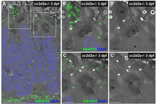
Fig. S10
SNAP25 mislocalizes in OSs and accumulated vesicles of cc2d2a-/- PRs at 3 dpf.
(A) 3 dpf correlative light and electron microscopy (CLEM) image of a cc2d2a-/- retina stained with anti-SNAP25 (green) and DAPI (blue, nuclei). (B-C?) Higher magnification images of the boxed regions in (A). SNAP25 mislocalizes in misshapen outer segments (B) and accumulated vesicles (C). (B? and C?) are SEM images only of (B and C). Arrows point to vesicular structures where SNAP25 mislocalizes. Scale bars: 4 ?m in A and 1 ?m in B-C?. OS outer segment, m mitochondria, N nucleus, P pigment, v vesicular structures.
Acknowledgments
This image is the copyrighted work of the attributed author or publisher, and
ZFIN has permission only to display this image to its users.
Additional permissions should be obtained from the applicable author or publisher of the image.
Full text @ PLoS Genet.