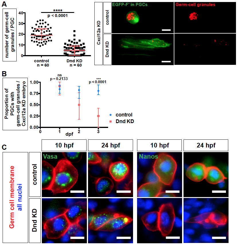
Fig. S4
Related to Figure 6. Germ cell granules in Dnd-deficient PGCs. (A) The number of germ cell granules per cell in Dnd-depleted and Cxcl12a knockdown embryos at 24 hpf (left). Maximum-intensity projection images of PGCs (Farnesylated EGFP, green) at 24 hpf showing germ cell granules (Granulito-dsRed, red) (right). Number of cells (n). Scale bars, 10 ?m. p-values were determined using the Mann-Whitney-U test with ?=0.05. Data are presented as median ± IQR. (B) Proportion of PGCs containing germ cell granules in Cxcl12a knockdown embryos upon depletion of Dnd between 1 and 3 dpf as compared to control treated PGCs. Number of embryos (18?N?38). Embryos lacking PGCs were omitted from the analysis. p-values were determined using the Mann-Whitney-U test with ?=0.05. Data are presented as median ± IQR. (C) Localization of the germ granule components Vasa and Nanos (green) in PGCs knocked down for Dnd (red membrane). EGFP fusion proteins of Vasa and Nanos translated from injected mRNAs that contained 3?UTR of the corresponding genes. 10 ?m thick confocal plane images captured at 10 and 24 hpf. Scale bars, 10 ?m. Farnesylated (F?), Knockdown (KD), hours post fertilization (hpf), days post fertilization (dpf).
Reprinted from Developmental Cell, 43, Gross-Thebing, T., Yigit, S., Pfeiffer, J., Reichman-Fried, M., Bandemer, J., Ruckert, C., Rathmer, C., Goudarzi, M., Stehling, M., Tarbashevich, K., Seggewiss, J., Raz, E., The Vertebrate Protein Dead End Maintains Primordial Germ Cell Fate by Inhibiting Somatic Differentiation, 704-715.e5, Copyright (2017) with permission from Elsevier. Full text @ Dev. Cell

