
|
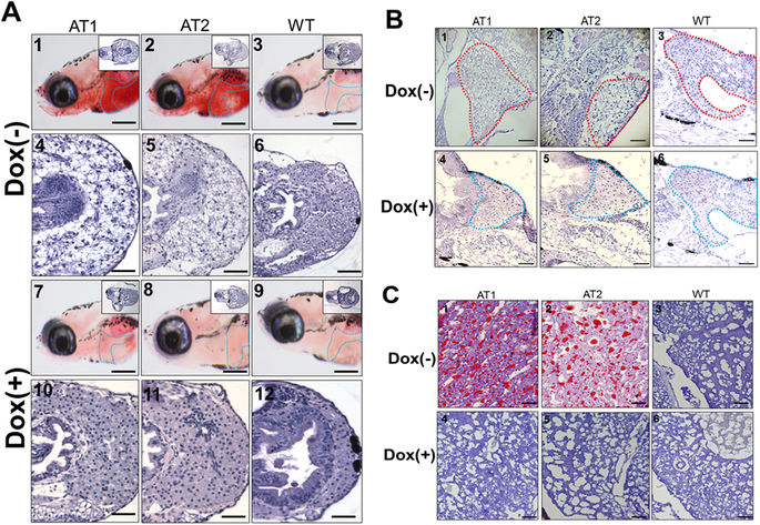
Fig. 4
Histological changes in livers of ATs???Dox larvae and juvenile ATs (<30 days post fertilization (dpf)) at different quantities of feeding and lipid contents. (A) Whole-mount ORO staining in the liver region of AT1?�?Dox (panels 1 and 7), AT2?�?Dox (panels 2 and 8), and WT?�?Dox (panels 3 and 9) larvae at 10 dpf (110X magnification, scale bars: 100 ?m). Livers are circled. H&E staining in the liver of AT1?�?Dox (panels 4 and 10), AT2?�?Dox (panels 5 and 11), and WT?�?Dox (panels 6 and 12) larvae. (400X magnification, scale bars: 200 ?m) (B) H&E staining in the liver region of juvenile (28 dpf) AT1?�?Dox (panels 1 and 4), AT2?�?Dox (panels 2 and 5), and WT?�?Dox (panels 3 and 6). Livers are circled. 200X magnification, scale bars: 200 ?m (C) Frozen section ORO staining (frozen ORO) in the liver of juvenile (28-dpf) AT1?�?Dox (panels 1 and 4), AT2?�?Dox (panels 2 and 5), and WT?�?Dox (panels 3 and 6). 400X magnification. Scale bars: 10 ?m.