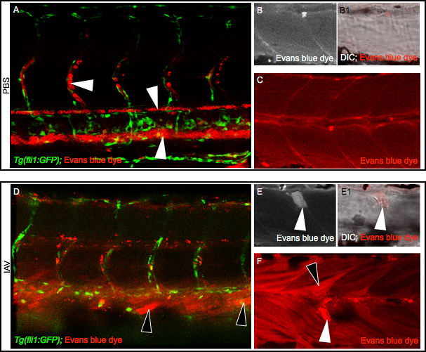
Fig. 2 IAV infection compromises the sarcolemma All embryo images are side mounts, dorsal top, anterior left. (A) Tg(fli1:GFP) zebrafish embryo with labeled endothelial cells (green) DC-injected with PBS plus EBD (red). EBD remains in the vasculature. White arrowheads point to EBD in an intersomitic vessel (top left), the dorsal aorta (middle), and the caudal vein (bottom right). (B-C) Wild-type zebrafish injected with PBS plus EBD. (B) Cropped EBD panel. (B1) Cropped EBD and brightfield panels merged. (C) EBD panel. Note that muscle fibers are impermeable to EBD in PBS-injected zebrafish. (D) Tg(fli1:GFP) zebrafish embryo DC-injected with IAV plus EBD (red). EBD leaked out of the vasculature and penetrated muscle fibers (black arrowheads). (E-F) Wild-type zebrafish injected with IAV plus EBD. (E) Cropped EBD panel. (E1) Cropped EBD and brightfield panels merged. (F) EBD panel. Note the uptake of EBD by long (black arrowheads) and retracted fibers (white arrowheads) indicative of sarcolemma damage.
Image
Figure Caption
Figure Data
Acknowledgments
This image is the copyrighted work of the attributed author or publisher, and
ZFIN has permission only to display this image to its users.
Additional permissions should be obtained from the applicable author or publisher of the image.
Full text @ PLoS Curr.

