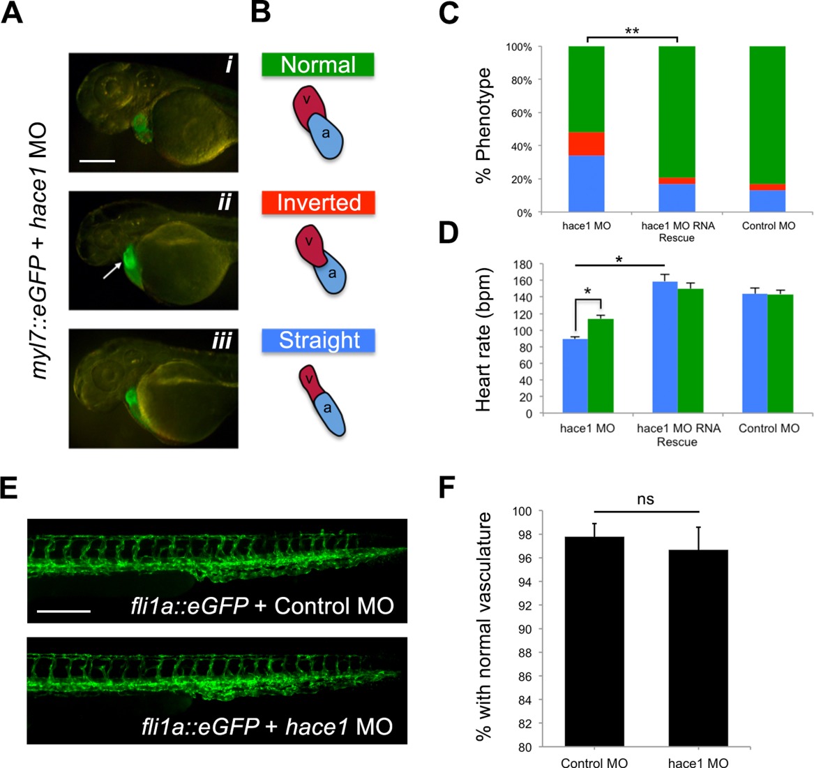
Fig. 2 hace1?morphant embryos display specific cardiac phenotypes that can be rescued by human HACE1 mRNA while the vasculature remains intact. A: The looping defect that results from hace1 knockdown results in either misalignment of the atrium and the ventricle (the ventricle is located on the left side [white arrow], ?inverted?) in ii or a tubular heart (?straight') in iii. Scale bar?=?250?Ám. B: Diagrammatic representation of each phenotype is included adjacent to a fluorescent image of a Tg(myl7::eGFP) hace1?morphant embryo with the associated phenotype. C: Bar graphs showing the percent of each of the representative anatomic phenotypes shown in panels A and B (colors correspond to the respective phenotypes; n?=?90?130 embryos for each group in 3 replicates; lateral views at 48 hpf; **P??0.05). Scale bar?=?250?Ám.
Image
Figure Caption
Figure Data
Acknowledgments
This image is the copyrighted work of the attributed author or publisher, and
ZFIN has permission only to display this image to its users.
Additional permissions should be obtained from the applicable author or publisher of the image.
Full text @ Dev. Dyn.

