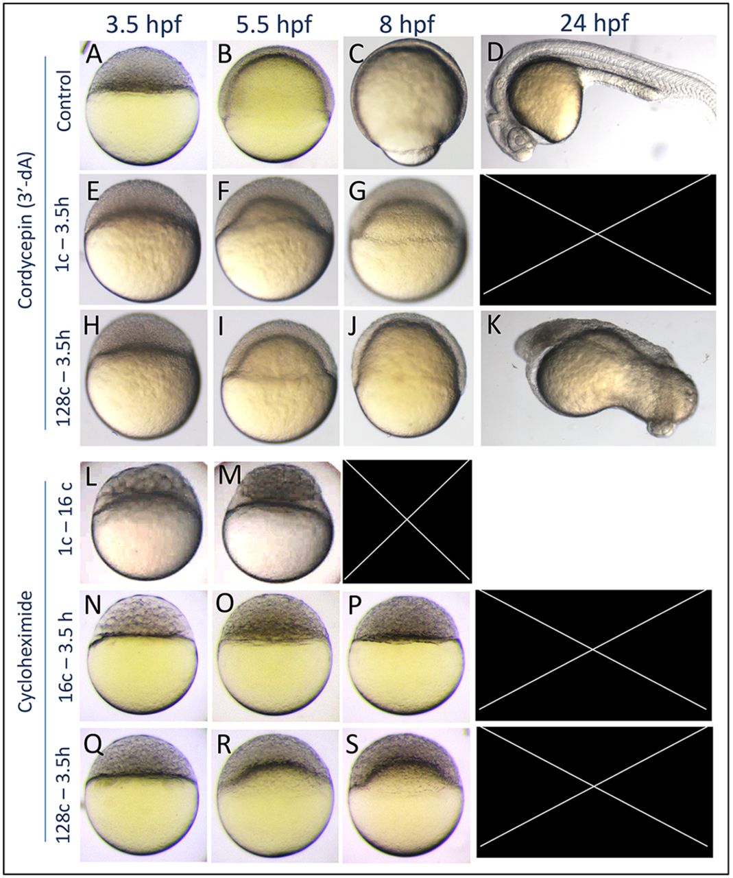
Fig. 3 Epiboly defects caused by 3?dA and CHX treatments. (A-D) Untreated control embryos. (E-G) Embryos that were treated with 3?-dA from the one-cell stage to 3.5?hpf undergo developmental arrest and cytolysis before 24?hpf (n=40). (H-K) 3?-dA treatment from the 128-cell stage to 3.5?hpf caused a delay of epiboly and gross patterning defects (n=30). (L,M) Translation inhibition by CHX treatment that was initiated at the one-cell stage affected early development and caused early lethality (n=20), whereas CHX treatment initiated at the 16-cell stage (N-P) resulted in developmental arrest at the oblong stage (n=61). Notice the larger cells at 3.5?hpf, suggesting defects in cell division. (Q-S) Treatment that was initiated at the 128-cell stage resulted in epiboly arrest followed by mortality (n=66).
Image
Figure Caption
Acknowledgments
This image is the copyrighted work of the attributed author or publisher, and
ZFIN has permission only to display this image to its users.
Additional permissions should be obtained from the applicable author or publisher of the image.
Full text @ Development

