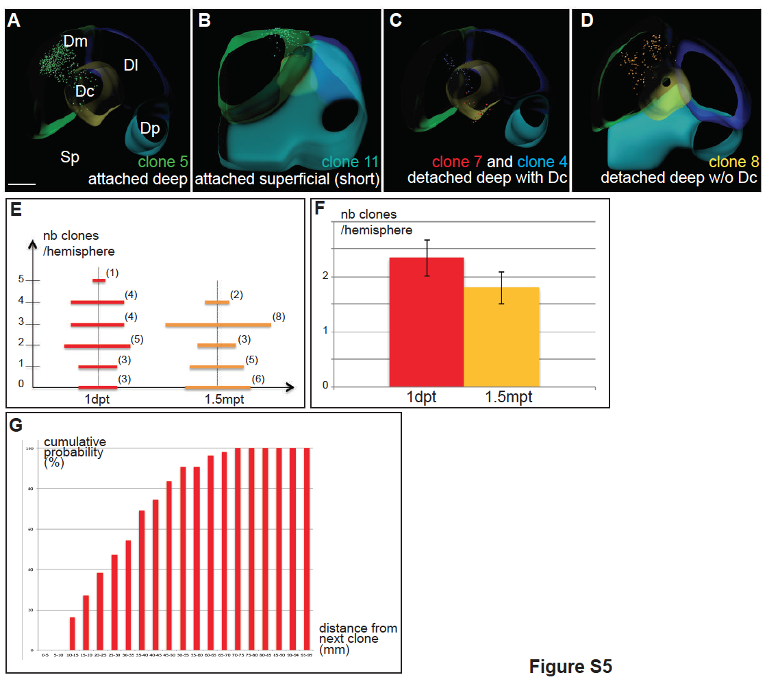
Fig. s5
Superimposed individual brainbow clones and neuroanatomical domains, related to Figs.5 and 6. A-D. 5 clones are shown on thick cross sections of the brain shown in Fig.5A. Clone categories, as defined in Fig.5 C-G and Table S1, are indicated at the bottom right of each panel, and the clones illustrated are color-coded as in Fig.5C-I and Table S1. Note that clone n°5 and 7, shown in A and C respectively, reach into Dc. Scale bar: 80?m. E-G. Validation of clonality in the analysis of her4actswitch,T(5dpf) larvae. E. Compared distribution of the number of induced progenitors at 1 day-post-9TB (1dpt) and the number of clones at 1.5mpf (1.5 months post 9TB ?mpt-). n = 20 pallial hemispheres at 1dpt and 24 pallial hemispheres at 1.5mpt. The numbers in bracket indicate the number of hemispheres concerned for each number of induced cells/clone. F. Average number of induced progenitors per hemisphere at 1dpt or induced clones per hemisphere at 1.5mpt (averaged from panel E). sem: 0.32 at 1dpt, 0.28 at 1.5mpt. Differenceis not significant (P<0.25). G. Cumulative distribution of the nearest neighbors distances separating labeled progenitors in the pallium of a her4H2a-mCherry,9TB(5dpf) animal analyzed at 1dpt (Fig.6A, A?). 84% of induced clones are located at least 15?m (approx. 4 cell diameters) away from their nearest neighbor clone.

