
Image
|
Figure Caption
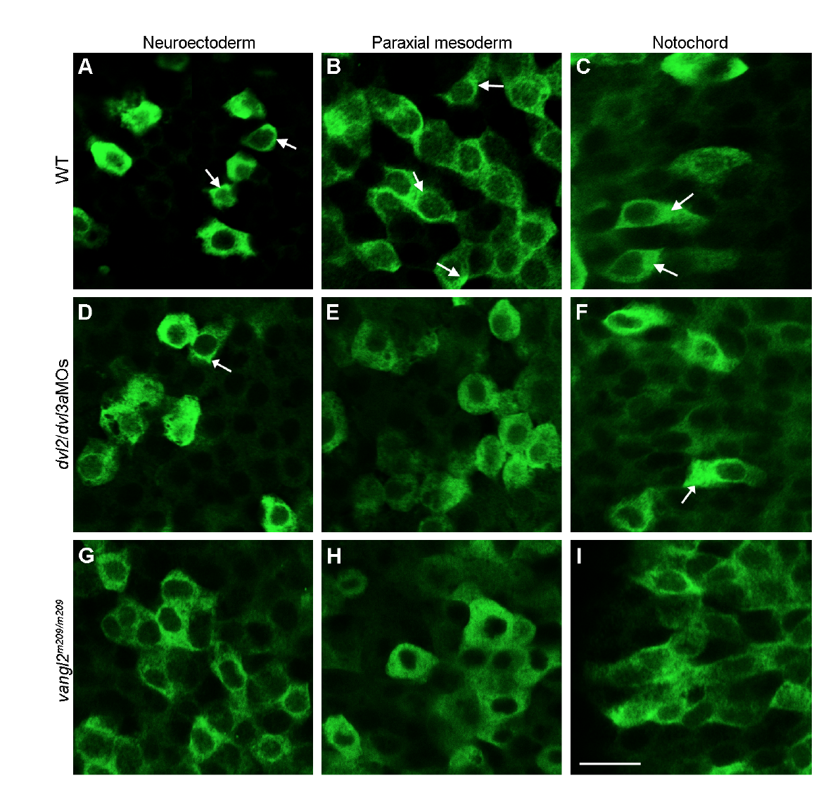
Fig. s10
Analysis by confocal microscopy of the subcellular localisation of myc-tagged Lurap1 in different germ layers at 80% epiboly stage, as indicated. (A-C) Subcellular localisation of Lurap1 in a WT embryo. Some cells (arrows) show localised distribution. (D-F) Subcellular localisation of Lurap1 in a dvl2 and dvl3a morphant embryo. (G-I) Subcellular localisation of Lurap1 in a zygotic vangl2m209/m209 mutant. Scale bar: (A-I) 20 μm.
Acknowledgments
This image is the copyrighted work of the attributed author or publisher, and
ZFIN has permission only to display this image to its users.
Additional permissions should be obtained from the applicable author or publisher of the image.
Full text @ Nat. Commun.