Fig. 2
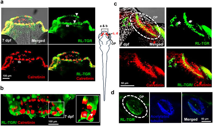
RL-TGR is expressed in the lip epithelium, taste buds, and olfactory epithelium. (a) Confocal images of the mouths of wild type embryos (dorsal view, anterior to the top) at 7 dpf stained with RL-TGR (green; expression in the epithelial layer of both upper and lower lip indicated by white arrowheads, basal cells and apical microvilli of the taste buds marked as ?tb?) and taste bud marker calretinin (red, expression in labial taste buds, also marked as ?tb?). (b) 3-D confocal images of the lips of 7 dpf wild type embryos (anterior of the lip facing outward) stained with RL-TGR (green) and calretinin (red). RL-TGR-positive cells were detected in the lip epithelium, basal cells, and apical microvilli (white arrowheads in the zoomed in inset) of taste buds (tb). (c) Confocal images of olfactory pit (OP) of wild type embryos (dorsal view, anterior to the top) stained with RL-TGR (green; expression in the sensory epithelium of the olfactory pit outlined by white dotted circles) and calretinin (red, expression in the olfactory receptor neurons of the olfactory epithelium). (d) Confocal images of olfactory pit (OP) of wild type embryos (dorsal view, anterior to the top) stained with RL-TGR (green; expression in the sensory epithelium of the olfactory pit outlined by white dotted circles) and acetylated tubulin (blue, expression in the cilia of the olfactory epithelium). n?>?20 embryos; schematic diagram of the 7 dpf larvae shows the location at 7 dpf of different images; the scale bars are shown in individual images.

