
|
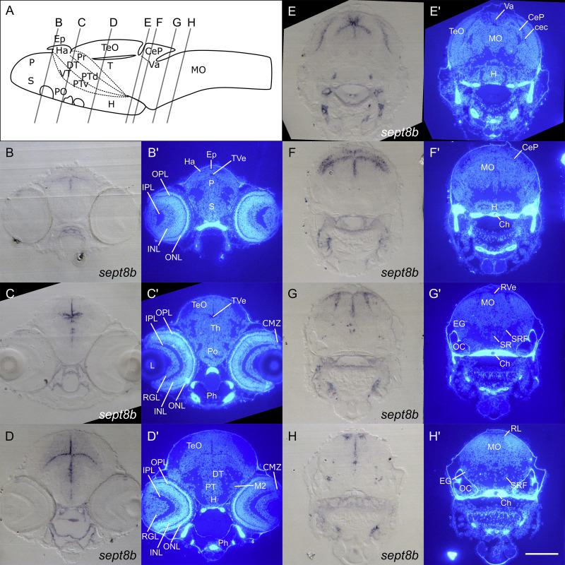
Fig. 6 sept8b expression in the CNS of 4 dpf zebrafish.
Images of transverse epon sections of 4 dpf zebrafish after labelling of sept8b transcripts via RNA in situ hybridization, counterstained with Hoechst33258 (B ? H). Levels and angle of sections are illustrated schematically in the drawing (A). Note that the illustrated scheme displays the structure of a 5 dpf zebrafish brain and, thus, can only be used for rough orientation. sept8b transcripts could be detected in ventricular cells of the forebrain and midbrain (B ? D), the Va and at border regions between TeO and CeP (E, F), in cells of the RL (G, H) as well as in accumulations of cells in the H (E) and the EG (H) and in single cells close to the SR (G). See list for abbreviations. Brain schemes were modified from M�ller and Wullimann (2016). Scale bars: B ? H: 100μm.
Reprinted from Gene expression patterns : GEP, 25-26, Berger, C., Helmprobst, F., Chapouton, P., Lillesaar, C., Stigloher, C., sept8a and sept8b mRNA expression in the developing and adult zebrafish, 8-21, Copyright (2017) with permission from Elsevier. Full text @ Gene Expr. Patterns