Fig. 5
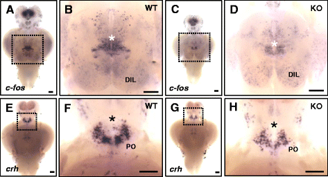
In situ hybridization of c-fos and crh in the brain of KO and WT fish. a?d Detection of c-fos mRNA expression in the dissected brain of WT (a, b) and KO (c, d) fish. Induction of strong c-fos expression is seen in specific brain regions (white asterisk) in WT fish (b) during social interaction, compared to that in KO fish (d). Higher expression of c-fos is observed in the diffuse nucleus of the inferior lobe (DIL) in the KO fish brain. fb, forebrain; mb, midbrain; hb, hindbrain; hy, hypothalamus. Ten animals for each WT and KO fish analysis were used. e?h Detection of crh expression in the brain of WT (e, f) and KO (g, h) fish. After acute social isolation for 24 h, crh-expressing cells are slightly reduced in preoptic area (PO, black asterisk) in KO fish (h), compared to that of WT fish (f). Eight animals for each WT and KO fish analysis were used. Anterior to the top and ventral view. b, d, f, and h Magnification of the ventral hypothalamic region boxed in a, c, e, and g. Scale bars 0.2 mm (a, c, e, and g), 0.76 mm (b, d), and 0.94 mm (f, h)

