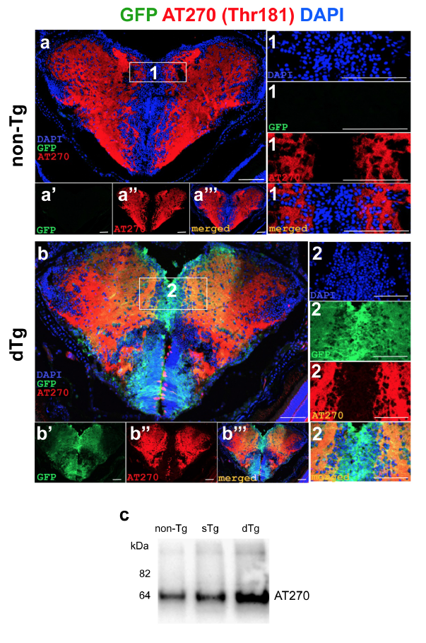
Fig. s8 (a) Immunohistochemistry (IHC) for AT270 (red) and GFP (green) on coronal sections of telencephalon of a 6-month old nontransgenic animal. (a?, a??) Individual fluorescent channels for GFP (a?) and AT270 (a??). Insets show the enlarged view of the frame in a with individual channels for DAPI, GFP and AT270, and merged image. (b) IHC for AT270 and GFP on coronal sections of telencephalon of a 6-month old dTg animal. (b?, b??) Individual fluorescent channels for GFP (b?) and AT270 (b??). Insets show the enlarged view of the frame in b with individual channels for DAPI, GFP and AT270, and merged image. (c) Western blot for AT270 from brains of non-Tg, sTg and dTg animals. Scale bars equal 50 ?m. n = 6 fish for every staining.
Image
Figure Caption
Acknowledgments
This image is the copyrighted work of the attributed author or publisher, and
ZFIN has permission only to display this image to its users.
Additional permissions should be obtained from the applicable author or publisher of the image.
Full text @ Sci. Rep.

