
|
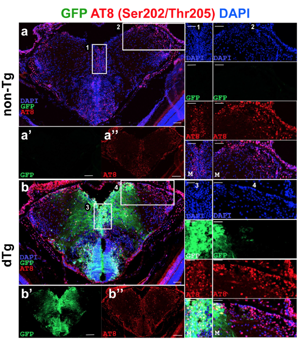
Fig. s5 (a) Immunohistochemistry (IHC) for AT8 (red) and GFP (green) on coronal sections of telencephalon of a 6 month-old non-transgenic animal. (a?, a??) Individual fluorescent channels for GFP (a?) and AT8 (a??). (1 and 2) The enlarged view of the inset in A with individual channels for DAPI, GFP and AT8, and merged image. (b) IHC for AT8 and GFP on coronal sections of telencephalon of a 6-month old dTg animal. (b?, b??) Individual fluorescent channels for GFP (b?) and AT8 (b??). (3 and 4) The enlarged view of the inset in b with individual channels for DAPI, GFP and AT8, and merged image. Scale bars equal 25 ?m. n = 5 fish for every staining.