
Image
|
Figure Caption
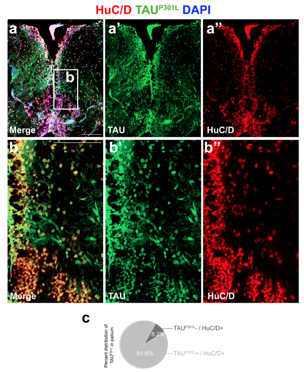
Fig. s4 Immunohistochemistry for HuC/D (red) and TAUP301L (green) on the telencephalon of a 6 month-old dTg animal. (a?, a??) Individual channels for green and red. (b-b??) High-magnification image of the frame in a. (c) Quantification of the percentage of HuC/D-positive neurons expressing TAU in the pallium. Scale bars equal 100 ?m. n = 4 fish and >20 histological sections for every staining and quantification.
Acknowledgments
This image is the copyrighted work of the attributed author or publisher, and
ZFIN has permission only to display this image to its users.
Additional permissions should be obtained from the applicable author or publisher of the image.
Full text @ Sci. Rep.